Video library
Colorectal health, explained
43 short videos on the conditions patients ask about most — what they are, why they happen, and what treatment actually involves. Recorded and reviewed by Dr. Kamrava.
Allen Kamrava, MD, MBA, FACS, FASCRS· Board-certified in Colon & Rectal Surgery and General Surgery· Reviewed
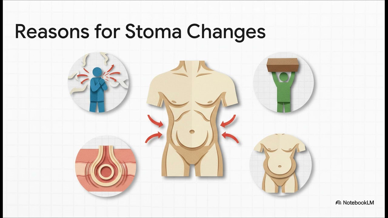 Stoma Changing Size: When It Pushes Out or Pulls InThis video explains two common changes in stoma size: prolapse and retraction. Stoma prolapse occurs when the bowel pushes out and the stoma appears longer. It is significantly more common in loop stomas (up to 47%) than in end stomas (up to 8%). Stoma retraction is the opposite, where the stoma pulls back in, sitting flat or below the skin, making a leak-proof seal difficult and risking skin irritation. Causes for both conditions are often related to anatomy or increased internal abdominal pressure (e.g., from coughing or lifting). Initial management focuses on home care. Retraction can often be managed with a convex pouching system, which gently pushes the stoma out. Prolapse can be managed with a support belt, and sometimes gently guided back in (reduced). Patients must call a doctor immediately if a prolapsed stoma appears dark, purplish, pale, cool, or very swollen and cannot be pushed back in, as this suggests a compromised blood supply. Surgery is considered the next step if home care fails or if the change causes blockages, pain, or major pouching problems.
 STOMA Skin Care: Stop Redness and LeaksThis guide focuses on managing paristomal dermatitis (skin irritation around the stoma), which affects up to 70% of people with a new stoma. The main cause is leakage of stoma output onto the skin, which creates a "vicious cycle" of irritation, moisture, and pouch failure. Two types of irritation are described: irritant dermatitis (red, raw, moist skin from output contact) and fungal infection (itchy, with small satellite spots). The most important defense is prevention through a perfect fit, ensuring the skin barrier snugly fits the stoma. Barrier rings can be used like caulk to fill gaps and create a level surface. For skin that is already moist or "weepy," stoma powder should be used to soak up moisture and secure the next appliance. Fungal infections require a topical antifungal powder (like Nystatin) followed by a skin sealant wipe to seal the powder in. The care routine involves cleaning, applying the correct powder (and brushing off excess), sealing the powder with a barrier wipe, using a barrier ring if needed, and applying the appliance. If skin issues persist after a couple of appliance changes, patients should consult their Wound Ostomy Incontinence Nurse (WOCN).
 When Your Stoma Is Too Fast: Simple Steps to Slow It DownThis content discusses a high output stoma (typically a small bowel stoma) defined as output exceeding 1,200 mL (1.2 L) in a 24-hour period. This rapid movement prevents the small intestine from adequately absorbing water and nutrients. High output is visually characterized by a thin, watery consistency (like "tea") compared to normal output (like "applesauce"). The primary risks are serious dehydration, electrolyte loss, weakness, and potential kidney problems. The most powerful defense is diet management. Patients should consume foods rich in soluble fiber (like bananas, bread, crackers, peanut butter) to soak up water and thicken the output. They should avoid sugary items (juice, soda) which draw more water into the intestine. Crucially, plain water should be avoided in favor of an Oral Rehydration Solution (ORS), which contains the perfect mix of salt and sugar to aid fluid absorption. If dietary changes are insufficient, the next step is medication, starting with Loperamide to slow the gut, sometimes followed by drugs that reduce stomach fluids. Medications should be timed about 30 minutes before eating. Warning signs of impending dehydration requiring immediate medical attention include feeling dizzy or lightheaded, having dark urine, extreme weakness, or muscle cramps.
 Leak After Bowel Surgery: What It Is and How It's TreatedThis explainer details the potential complication of an anastomotic leak following bowel surgery. The goal of surgery is to create a strong, watertight connection (anastomosis) using healthy tissue, excellent blood supply, and zero tension. A leak occurs when the delicate healing balance is disrupted, and a gap forms in the new seal, allowing bowel contents to escape. The risk ranges from 2% to 21% of cases. Nearly half of all leaks can appear after the patient has gone home. Key warning signs include fever, abdominal pain that is worsening (not improving), unusual fluid from the incision, or a generalized feeling of being unwell. Treatment is personalized. Smaller leaks in stable patients might be managed non-surgically with antibiotics and potentially a drain. Larger leaks or if the patient is very sick may necessitate a return to the operating room. Surgeons may create a temporary stoma (a detour) to route stool away from the healing connection, allowing it to rest and heal properly. Scarring from a leak can sometimes lead to long-term changes in bowel function.
 Bleeding After Surgery: What's Normal and What's an Emergency?This content provides straightforward information regarding post-operative bleeding, defined as any bleeding that happens after an operation. It is emphasized that this is a known risk for which medical teams are always prepared. Factors that can increase the risk of bleeding include the location of the surgery (e.g., right side of the colon), the use of blood thinners, removal of a large polyp, and a patient's overall health. Crucially, the video teaches viewers to distinguish between normal healing (a little spot of blood on a bandage, bruising) and signs that require immediate medical attention. Emergency signs include feeling dizzy, weak, or lightheaded, having a very fast heart rate, or seeing heavy bleeding that does not stop. Management is tailored to the severity. For mild bleeding, monitoring or medication adjustment may suffice. For significant bleeding, doctors often use endoscopic tools (scopes) to apply tiny clips or use heat to seal things up without new cuts, a technique that has an incredible success rate of 95% to 100% for bleeding after a colonoscopy/polyp removal. Returning to the operating room is reserved for serious cases. Patients are urged to be active partners in their recovery by following medication instructions and reporting any new or worsening symptoms right away.
 Bulge Near Your STOMA: What Is a Parastomal Hernia?This content serves as a guide to understanding a parastomal hernia, which is a new bulge around the stoma. The abdominal wall acts like fabric, and the hernia forms when the surgical opening for the stoma stretches and weakens, allowing internal matter (intestine or fatty tissue) to push through. Parastomal hernias are extremely common, developing in over 50% of people with a stoma long-term. Symptoms that prompt medical consultation include pain, difficulty getting the pouch to stick (leading to leaks/irritation), or feeling like output is blocked. End colostomies carry the highest risk, while loop ileostomies have the lowest. A major predictor of risk is body shape; having a waistline over 100 cm (about 40 inches) is linked to a staggering 75% chance. Other risk factors include being overweight, age, diabetes, smoking, and emergency surgery. Treatment involves a personalized decision between watchful waiting (chosen by up to 70% of people) or surgical repair. If surgery is chosen, pre-operative optimization is critical, requiring measures like achieving a BMI under 35 and being smoke-free for at least 4 weeks. Surgical repair involves using a special mesh, as stitching the hole closed fails up to 80% of the time. The preferred technique is the minimally invasive Sugarbaker method, which guides the intestine out from underneath the edge of the mesh to create a one-way valve effect. The Sugarbaker method has a recurrence rate around 10%, significantly lower than older methods. Despite effective repairs, long-term recurrence rates can still climb over 50%.
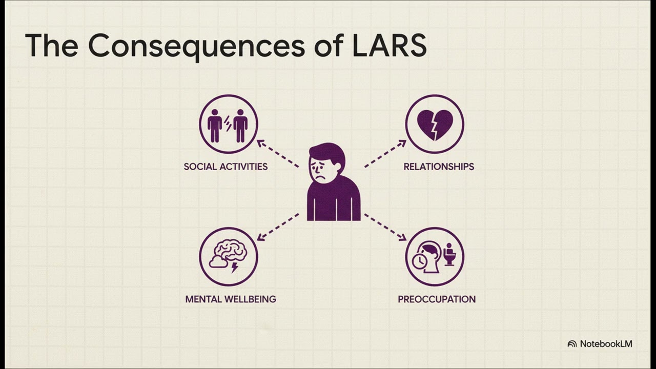 LARS Explained: Bowel Problems After Rectal SurgeryThis video addresses Low Anterior Resection Syndrome (LARS), a set of challenges experienced following rectal surgery that can be long-lasting and is considered a chronic condition. LARS results from the surgery changing the "internal plumbing," impacting sensitive nerves, pelvic floor muscles, and removing the rectum (the body's natural storage tank for stool). LARS is defined as disordered bowel function after rectal resection that leads to a detriment in quality of life. Symptoms include urgency, incontinence (soiling), frequent bowel movements, the sensation of incomplete emptying, and clustering (multiple movements in a short period). Diagnosis is usually confirmed about 3 months after surgery or ileostomy reversal, often starting with the patient keeping a bowel diary. Treatment is viewed as building a personal toolkit, following an additive, step-by-step strategy ("treatment ladder"). The foundation involves diet, fiber, and hydration adjustments, followed by medications to regulate function. A key procedure is retrograde colonic irrigation (RCI), a specific technique using water to gently wash out the colon on a predictable schedule, allowing for long symptom-free periods. The goal of management is not a perfect cure, but rather meaningful improvement in quality of life.
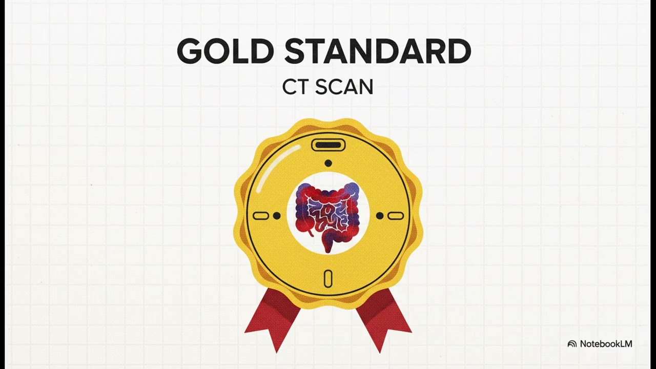
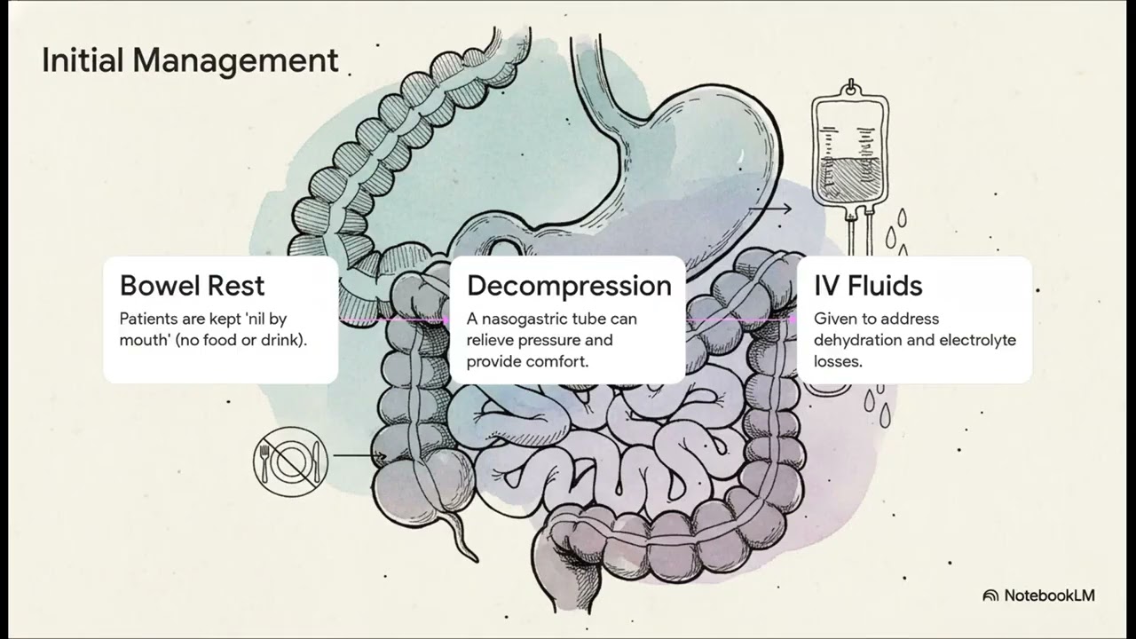 Digestive Traffic Jam: Understanding a Blocked BowelThis video explains a bowel obstruction as a "traffic jam" inside the digestive system where a blockage stops the flow of gas, fluid, and food, causing the bowel to swell. Causes are grouped into three categories (inside the intestine, in the wall, or from outside pressure). The focus is on the single biggest cause of small bowel obstructions: scar tissue or adhesions (sticky bands forming after abdominal surgery) that twist or squeeze the intestine. Classic warning signs include abdominal distension (swelling, seen in 2/3 of patients), abdominal pain (starting as cramping waves and becoming constant), nausea, vomiting, and most notably, the inability to pass gas (90% of people) or have a bowel movement (over 80% of people). Diagnosis involves a physical exam, medical history (especially past surgeries), and imaging. While X-rays can show swollen loops of intestine, the CT scan is the gold standard because it provides a detailed 3D picture showing the exact location and cause of the blockage, with an accuracy of about 96% for large bowel obstructions. Initial treatment aims to stabilize the patient by resting the bowel (stopping eating/drinking), draining backed-up fluid and air with an NG tube, and providing IV fluids. For obstructions caused by adhesions, the common starting point is non-surgical management, which is highly successful, with rates reaching 87%. Surgery is only pursued if the non-surgical approach fails or if there are signs of a more serious problem.
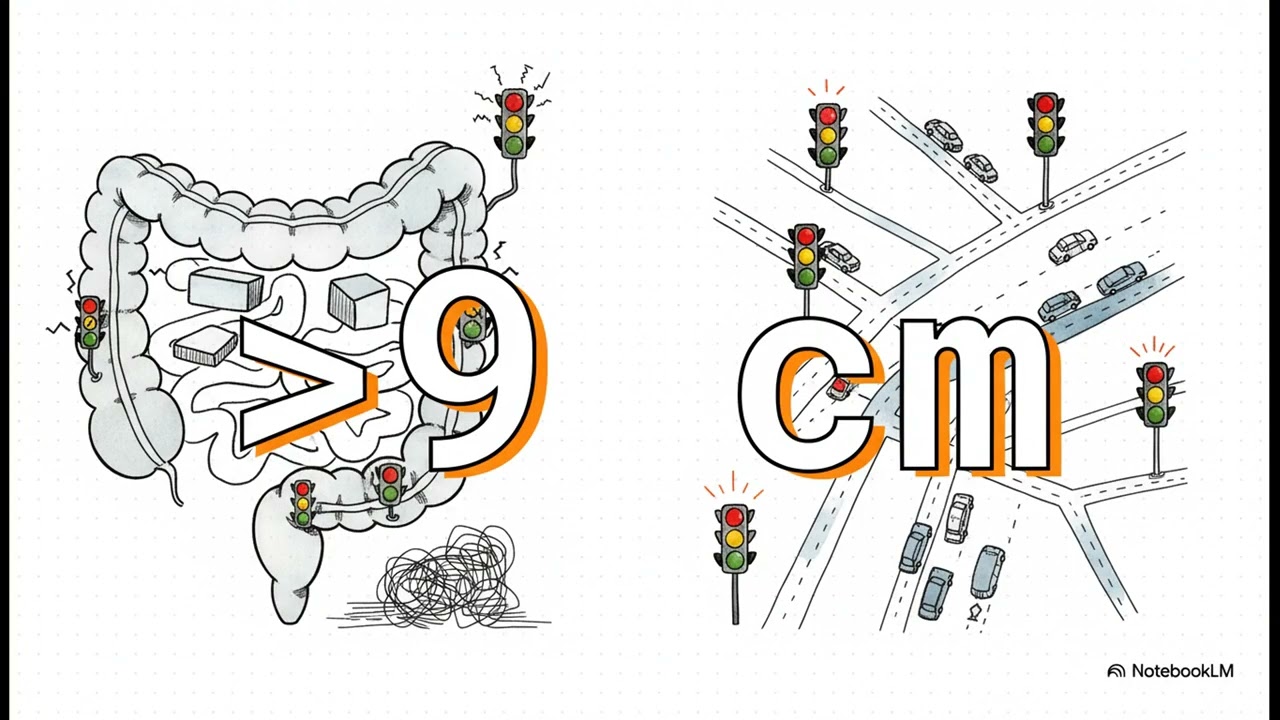 "False" Blockage: When Your Bowel's Signals Break DownThis video explains acute colonic pseudo obstruction, a condition where the colon acts blocked but there is no physical obstruction (a "false" blockage, as "pseudo" means false). It is described using the analogy of a clear highway where the traffic signals (nerve signals) are broken, preventing movement. This condition rarely appears on its own; it is typically a body reaction to major stress, such as recovering from a big surgery or a tough infection. Diagnosis relies on imaging (X-rays or CT scans) to show the colon is dilated (widened) while confirming there is no actual mechanical obstruction. Close monitoring is essential, particularly tracking the size of the cecum (the widest part of the colon, which is under the most strain), with a measurement exceeding 9 cm being a major flag. If the cecum stretches past 10 or 12 cm, the risk of perforation (a tear) increases significantly, which is the main outcome doctors strive to prevent. Treatment follows a clear step-by-step process starting with the least invasive methods. Step one is supportive care (bowel rest, no food/drink, walking), which resolves the issue for an amazing amount of people (around 87%). If rest fails, Step two may involve the medication neostigmine to "jump start" the nerves. Step three is colonoscopic decompression (using a flexible scope to let the air out). Surgery is the absolute last resort reserved for very rare cases where perforation has occurred or all other options have failed.
 The Twisted Bowel: What You Need to KnowThis content breaks down volvulus (twisted bowel), a condition where the twist creates a total roadblock, trapping gas and fluid, and potentially cutting off the blood supply to the tissue. The location of the twist determines the treatment plan.
This is the more common type (60% of cases), occurring in the S-shaped colon, typically in older men. Symptoms include major abdominal swelling (distension), pain, and inability to pass gas or have a bowel movement. Diagnosis involves X-rays (looking for the "coffee bean sign" or "bird's beak sign" with dye) and confirmation via CT scan, which is nearly 100% accurate. Initial relief is provided by endoscopic detorsion (using a scope to untwist the bowel). However, because the risk of recurrence is high (45–70%), planned surgery to remove the floppy colon section is the standard permanent recommendation.
This type occurs where the small and large intestines meet, is less common, and is often seen in younger women. This is a surgical emergency as endoscopic detorsion is ineffective. The standard of care requires immediate surgery, specifically a right hemicolectomy, to remove the twisted section and reconnect the healthy ends.
The Twisted Bowel: What You Need to KnowThis content breaks down volvulus (twisted bowel), a condition where the twist creates a total roadblock, trapping gas and fluid, and potentially cutting off the blood supply to the tissue. The location of the twist determines the treatment plan.
This is the more common type (60% of cases), occurring in the S-shaped colon, typically in older men. Symptoms include major abdominal swelling (distension), pain, and inability to pass gas or have a bowel movement. Diagnosis involves X-rays (looking for the "coffee bean sign" or "bird's beak sign" with dye) and confirmation via CT scan, which is nearly 100% accurate. Initial relief is provided by endoscopic detorsion (using a scope to untwist the bowel). However, because the risk of recurrence is high (45–70%), planned surgery to remove the floppy colon section is the standard permanent recommendation.
This type occurs where the small and large intestines meet, is less common, and is often seen in younger women. This is a surgical emergency as endoscopic detorsion is ineffective. The standard of care requires immediate surgery, specifically a right hemicolectomy, to remove the twisted section and reconnect the healthy ends. Rectal Prolapse GuideRectal Prolapse occurs when the full thickness of the rectum wall turns inside out and sometimes sticks out through the anus. The condition can be internal (the rectum telescopes into itself) or external. Symptoms include a feeling of pressure or bulge, incomplete evacuation, mucus/stool leakage, and chronic straining. It is linked to the weakening of pelvic floor muscles and ligaments, often caused by chronic constipation, childbirth, or genetics. Diagnosis involves physical exam and specialized imaging, such as defecography, which provides a real-time view of the rectum's function during a bowel movement. Treatment is primarily surgical and is tailored to the patient. Surgical paths include the perineal approach (from below, higher recurrence, often for older/frail patients) and the abdominal approach (lower recurrence, often minimally invasive, such as ventral mesh rectopexy). The source notes there is no single "gold standard" operation.
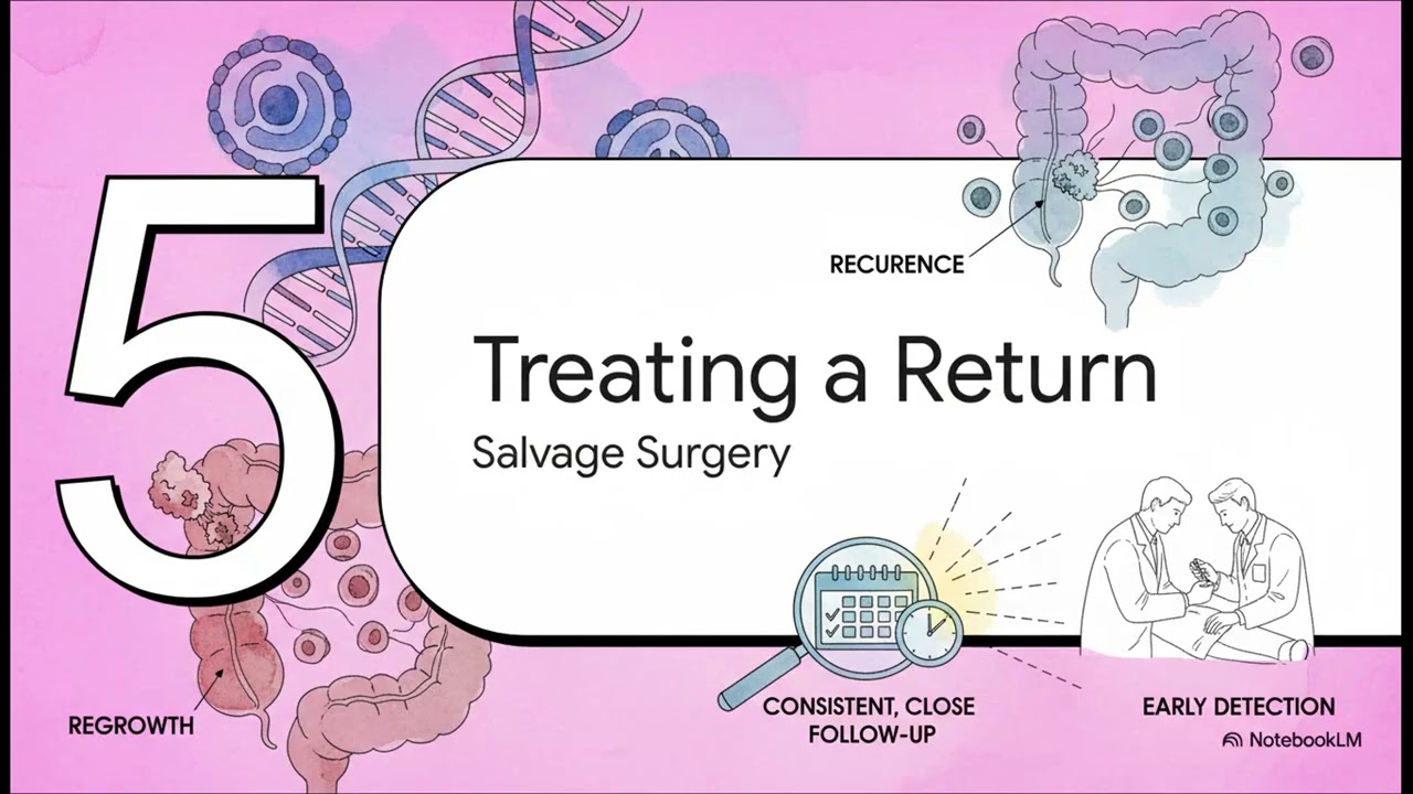 If Rectal Cancer Comes Back: What Happens Next?The content discusses the possibilities if rectal cancer returns after initial treatments like chemotherapy and radiation. The patient's path depends on the initial response, either "watch and wait" (if the tumor had a complete clinical response or CCR) or traditional surgery. If cancer returns after "watch and wait," it is called local regrowth (occurring in about 25% of candidates). If it returns after surgery, it is called a local recurrence; the rate of recurrence has plummeted to about 6% today due to advances like total mesorectal excision (TME). Consistent, close follow-up (surveillance) is the most critical element. If a return is found early, treatments are highly effective; for patients with local regrowth, salvage surgery can successfully remove the tumor and offer a cure about 90% of the time.
 SRUS (Solitary Rectal Ulcer Syndrome): Causes and Non-Surgical FixesSolitary Rectal Ulcer Syndrome (SRUS) is a benign (non-cancerous) disorder tied to the physical mechanics of defecation. Symptoms include bleeding, pain, straining, or tenesmus. The cause is mechanical: repeated straining causes the upper rectum to fold down into the lower part (internal telescoping), leading to friction and ulcers or irritated tissue. A definitive diagnosis requires an endoscopy and a biopsy, which identifies specific microscopic scar tissue in the rectal wall. For the vast majority of patients, the treatment is non-surgical. The goal is to fix the mechanical root cause by reducing straining through diet changes, bowel retraining, and pelvic floor physical therapy. Surgery is rarely the primary solution, reserved only for severe structural issues like a deep internal prolapse that cannot be fixed otherwise.
 What is a Rectal Neuroendocrine Tumor? Understanding Slow-Growing TumorsThe content focuses on rectal neuroendocrine tumors (rectal NETs), which are slow growing (indolent) growths starting in neuroendocrine cells. They are often found incidentally during routine colonoscopy, as most patients have no symptoms. Although sometimes called "carcinoid" tumors, their behavior is typically much less aggressive than standard colon cancer. Size is the most important factor for determining treatment and prognosis. Small tumors (under 1 cm) have an excellent prognosis (near 100% 5-year survival rate) and are usually removed during endoscopy. Medium tumors (1–2 cm) are usually treated with local excision, and large tumors (over 2 cm) require more formal surgery.
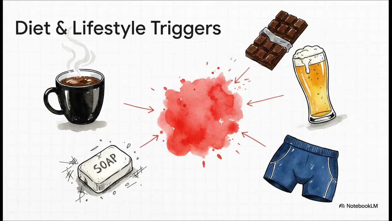 Anal Itching (Pruritus Ani): Breaking the Itch-Scratch CycleThis guide discusses Pruritus Ani (anal itching), a common medical issue affecting up to 5% of the general population. The core concept explored is the itch-scratch cycle, where scratching causes injury and inflammation, leading to a worse itch. Causes can often be found (75% of the time) and relate to lifestyle (coffee, chocolate, harsh soaps, tight fabrics) or conditions that cause moisture/stool leakage (chronic diarrhea, hemorrhoids). Diagnosis involves a conversation, visual check, and sometimes an anoscopy. Treatment focuses on breaking the cycle and creating a healing environment through simple changes: gentle cleaning (using only water), patting dry, increasing fiber, and wearing breathable cotton underwear. If lifestyle changes are insufficient, a doctor may recommend barrier creams, mild steroid creams, or antihistamines.
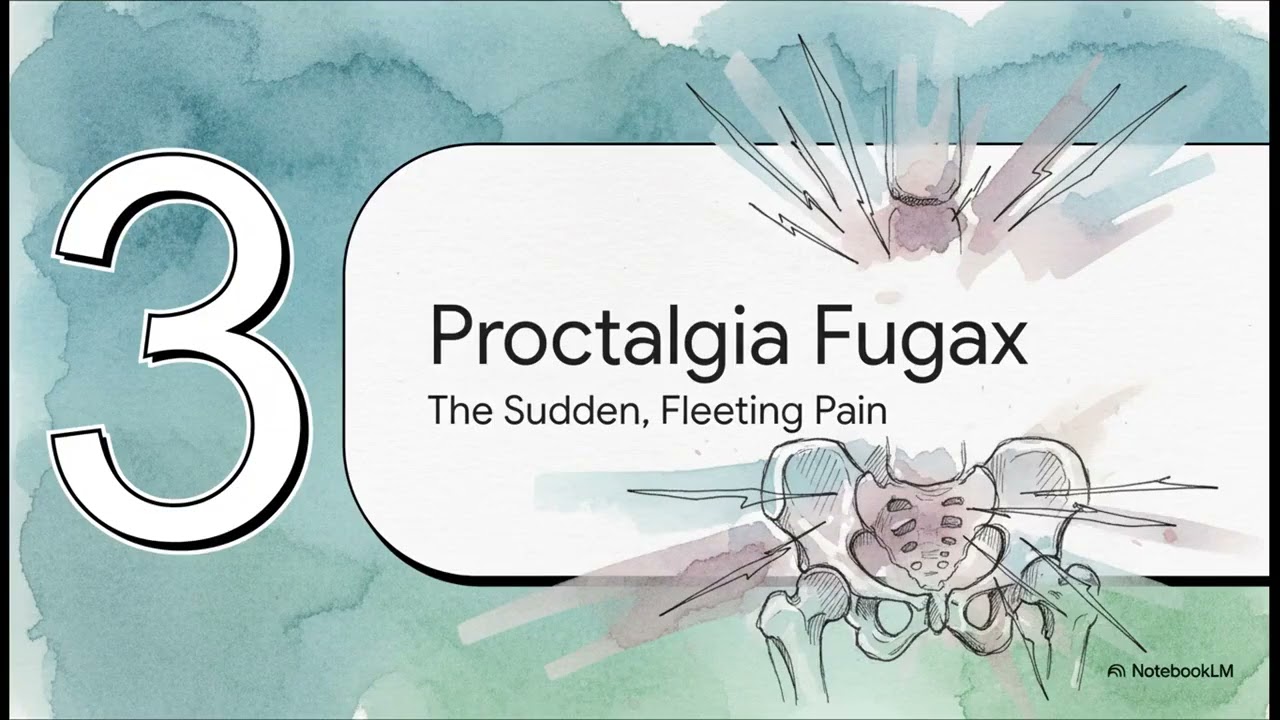 Deep Rectal Pain: Functional Causes (Spasms) & TreatmentThis content addresses functional anorectal pain, meaning pain caused by the function of muscles and nerves rather than a visible physical problem. The pain is often linked to the pelvic floor muscles, particularly the U-shaped puborectalis muscle. Two main types are identified based on duration: Levator Ani Syndrome (dull, persistent ache lasting over 30 minutes, often linked to chronically tight muscles and stress) and Proctalgia Fugax (sudden, sharp, fleeting pain lasting under 30 minutes, caused by anal sphincter or nerve spasms). Treatment for Levator Ani Syndrome focuses on muscle relaxation using biofeedback (conscious control training), physical therapy, or sitz baths. For Proctalgia Fugax, treatment involves reassurance (understanding the pain is not dangerous), warm baths, or specific medications like topical creams or albuterol inhalers to stop attacks.
 Pilonidal Disease: Causes, Symptoms & Treatment GuideThis content defines Pilonidal Disease (known during WWII as "Jeep disease") as an acquired condition, not congenital. It occurs when loose hair gets trapped under the skin in the natal cleft (crease at the top of the buttocks), which the body walls off, leading to inflammation, cysts, and potential abscesses. Risk factors include a deep natal cleft, coarse hair, sitting for long periods, obesity, and family history. Diagnosis is typically made via a simple physical exam. Treatment is individualized and patient-focused. Options range from conservative management (keeping the area clean/dry, hair removal, avoiding prolonged sitting) to surgical interventions. Surgical trade-offs exist between the lay open procedure (low recurrence, slow healing) and excision and closure (faster healing, higher recurrence). Advanced techniques, such as flap procedures (e.g., Cleft Lift), are used for recurrent cases to flatten the cleft's anatomy. Recurrence rates can be up to 34% after one year, making long-term hair removal and lifestyle changes vital.
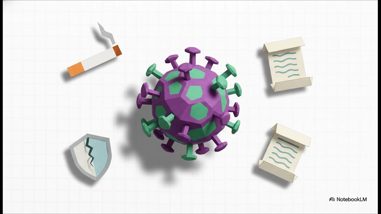 Anal Cancer: Modern Treatment & Favorable OutlookThis explainer provides an overview of anal cancer, defining it as cancer starting in the anus, differentiated by location (anal canal vs. perianal area). Approximately 85% of cases are Squamous Cell Carcinoma. The Human Papillomavirus (HPV) is considered the major driver behind most cases. Diagnosis involves a physical exam, biopsy (the only way to know for sure), imaging scans (CT/PET), and staging (TNM system: Tumor, Nodes, Metastasis). The modern standard of care is combination chemo-radiotherapy, which is highly successful and means that major surgery is typically avoided for the vast majority of patients. Post-treatment, careful monitoring is required for 5 years. The outlook is described as "imminently curable" with "favorable outcomes".
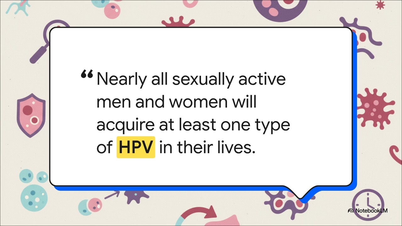 Abnormal Cells and HPV: Understanding AINThis video explains Anal Intraepithelial Neoplasia (AIN), a condition where cells in the anal canal lining appear abnormal. It is defined as a precancerous condition and is very similar to cervical dysplasia. AIN is caused by the Human Papillomavirus (HPV), particularly high-risk types like HPV 16 and 18. Cell changes exist on a spectrum, classified as low-grade lesions (LSIL) or high-grade lesions (HSIL), with doctors primarily focusing on treating HSIL to prevent progression to cancer. Diagnosis starts with anal cytology (similar to a Pap smear). If abnormal cells are found, a High-Resolution Anoscopy (HRA) and biopsy are performed for a definitive diagnosis. Treatment options are personalized: low-grade AIN often involves observation, while high-grade AIN is treated by removing abnormal cells using topical or ablative therapy. Long-term surveillance and follow-up are essential due to the potential for recurrence. The HPV vaccine is highlighted as a powerful tool for prevention.
 Understanding and Treating Crohn's Disease in the Anal AreaThis guide focuses on Perianal Crohn's disease, which affects 13% to 38% of people with Crohn's. Symptoms include persistent pain, swelling, drainage, and bleeding. Diagnosis utilizes physical exam, MRI (to map the internal structure), and sometimes an Exam Under Anesthesia (EUA) for thorough checking without pain. Treatment follows the "golden rule": eliminating active infection first. Abscesses (painful pockets of pus) require quick surgical drainage for pain relief and infection control. Fistulas (small tunnels) are treated with a combined approach: medical therapy using Biologics (Anti-TNF drugs) to address the underlying inflammation, and surgical placement of a seton (a soft drain) to allow for clean, slow healing and prevent recurrence. Other associated issues (fissures, ulcers) are usually managed by treating the underlying Crohn's. The overall treatment goal is eliminating pain and infection while preserving normal bowel function and improving the patient’s quality of life.
 Fecal Incontinence: Stop the Embarrassment and Find TreatmentThe video addresses fecal incontinence, described as a common and distressing medical condition that may affect up to 1 in 5 people. Continence is explained as a "team effort" involving stool consistency, sphincter strength, rectal capacity, and nerve signals. Causes include injury to sphincter muscles (e.g., childbirth), nerve issues (diabetes, spinal cord injury), chronic diarrhea, or radiation therapy. Diagnosis includes a conversation, physical exam, and specialized tests like an anal ultrasound. Treatment is staged, starting with non-invasive options: dietary changes (reducing caffeine), fiber supplements, anti-diarrheal medication, bowel programs, and biofeedback (physical therapy for the pelvic floor). Advanced treatments include Sacral Neuromodulation (SNM) (a device likened to a pacemaker for the bowel), injection of bulking agents to tighten the canal, or sphincteroplasty to repair torn muscles. For patients who have tried everything, ostomy surgery is presented as a means to regain control and quality of life.
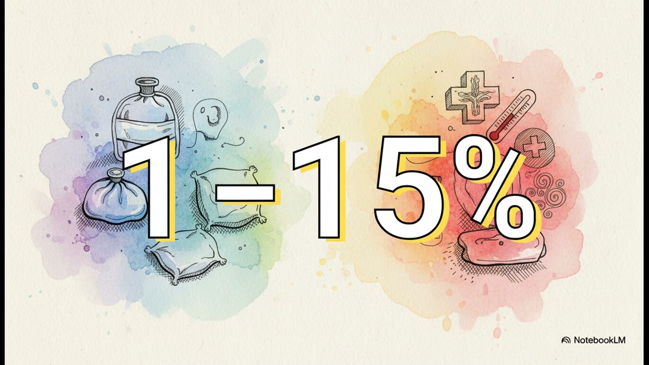 Smooth Recovery: A Simple Guide to Hemorrhoid SurgeryThis video guides patients through recovery after hemorrhoid surgery, focusing on pain management and healing. Pain is manageable using a layered approach: long-acting local anesthetic used during surgery, followed by non-narcotic medications like ibuprofen at home, with stronger pain meds reserved for breakthrough pain. Warm sitz baths are highly soothing. Preparing for the first bowel movement is critical, requiring significant hydration (at least 64 ounces of water daily) and fiber supplements to ensure soft stool, and the use of a footstool is recommended. Potential, usually temporary, complications include urinary retention (1% to 15% of patients). Patients are advised to distinguish between normal spotting (common) and heavy bleeding or passing blood clots (requires immediate medical attention). The surgery has "excellent results," with surgeons actively working to prevent scarring and narrowing of the anal canal by leaving "bridges of normal tissue".
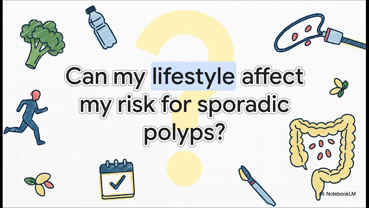 Colon Polyps: Good News, Not Bad NewsThis explainer clarifies that a colon polyp, or adenoma, is simply a small growth on the inside lining of the colon and is not cancer. Polyps form through a slow journey (taking a decade or longer) initiated by genetic changes or "typos" in the cell's DNA, which can cause cells to grow unchecked. Most colorectal cancers (about 80%) are sporadic, resulting from random genetic mistakes over a lifetime, rather than inherited genes. Lifestyle factors, such as high intake of red/processed meats or alcohol and lack of physical activity, can increase risk, while whole grains, dairy, and regular activity are protective. Screening, ideally starting at age 45 for average-risk individuals, is the solution. The colonoscopy is the best tool because it allows doctors to remove the polyp (polypectomy) immediately, thereby actively preventing potential cancer.
 Colorectal Cancer Basics: Prevention, Screening, and TreatmentThis guide covers the essentials of colorectal cancer, which is the third most common cancer globally, emphasizing that it is often preventable and highly treatable if caught early. Colorectal cancer is a cancer that begins in the large intestine (colon and rectum) when errors (mutations) in the genes cause cells to grow and divide out of control. Most cases (about 80%) are sporadic, while approximately 20% are inherited. The cancer typically begins as a benign growth called a polyp, and the slow progression from polyp to cancer (which takes several years) offers a critical "window of opportunity" for screening and prevention. Current recommendations suggest starting screening at age 45 for people at average risk. Treatment is personalized based on the cancer stage and genetic makeup, utilizing tools such as surgery (the cornerstone), chemotherapy, targeted therapy, and immunotherapy.
 Hereditary Non-Polyposis Colorectal Cancer (Lynch Syndrome): Understanding Your Family's Cancer RiskThis explainer details Lynch syndrome, which is the most common type of hereditary colorectal cancer, though it accounts for only about 3% of all colorectal cancer cases. It is a genetic condition that increases cancer risk. The root cause is a glitch in a "spell checker" gene within the mismatch repair (MMR) system, allowing genetic typos to accumulate and eventually leading to cancer. Current guidelines recommend universal screening (testing the tumor tissue) for everyone diagnosed with colorectal cancer to identify the faulty gene. Lynch syndrome carries the highest risk for colorectal cancer and the second highest risk for endometrial/uterine cancer in women. Management involves aggressive, proactive surveillance: colonoscopies start in the early 20s and are performed every 1 to 2 years, which can significantly improve life expectancy. Lynch-related cancers are often highly susceptible to modern immunotherapy drugs.
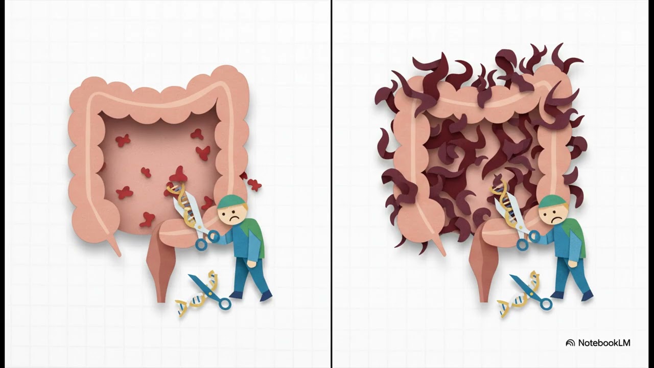 Familial Adenomatous Polyposis (FAP): Managing Hundreds of Colon GrowthsThe content explains Familial Adenomatous Polyposis (FAP), a rare inherited condition defined by the presence of more than 100 polyps (adenomas) in the colon and rectum, often starting at a young age. FAP is caused by a mutation in the APC gene, which normally controls cell growth. If the condition is not managed proactively, the risk of developing colorectal cancer is nearly 100%. While 75% of cases are inherited, 25% occur as a brand new mutation. Proactive management is crucial, involving colonoscopies starting around puberty and upper endoscopies later. The most important step is preventative surgery to remove the colon and/or rectum to stop cancer before it develops. Due to the widespread effect of the APC gene mutation, FAP can also lead to polyps in the stomach/small intestine, a higher risk for thyroid cancer, and serious, non-cancerous desmoid tumors.
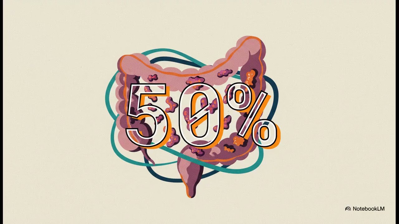 Serrated Polyposis Syndrome (SPS): Managing Many Serrated PolypsThis video explains Serrated Polyposis Syndrome (SPS), the most common type of polyposis syndrome. The name describes the condition of having a "bunch of polyps" that look jagged or "serrated" under a microscope. Diagnosis relies on specific WHO criteria, such as the "5 and 2 rule" or the "20 plus rule" (a lifetime count of more than 20 serrated polyps). SPS carries a lifetime risk of advanced, potentially cancerous growths up to 50%. However, this risk is drastically lowered by the management plan, which involves intensive surveillance (colonoscopies every 1 to 3 years) following initial clearance of all visible polyps. Key actions for patients include quitting smoking, as there is a very strong link between smoking and SPS, and informing family members about the need for screening.
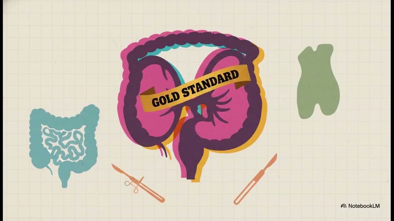 Ulcerative Colitis: When to Consider Surgery (J-Pouch Explained)This guide focuses on Ulcerative Colitis (UC), an IBD where inflammation is confined to the innermost lining (mucosa) of the large intestine. Surgery is a common, proactive option for about 3 out of 10 people with UC during their lifetime. Reasons for elective surgery include medication failure, intolerable side effects, or managing the increased risk of colorectal cancer (which jumps to 18% after 30 years with UC). The gold standard procedure is the J-pouch (Ileoanal Pouch Anastomosis or IPAA), which involves 1) removing the diseased colon/rectum, 2) folding the small intestine (ilium) into a J-shaped pouch, and 3) reconnecting the pouch to the anus to restore the natural pathway. It is emphasized that UC patients experiencing worsening symptoms must always be checked for a separate infection, such as C. diff or CMV, as mistakenly treating an infection with immunosuppressants can be dangerous.
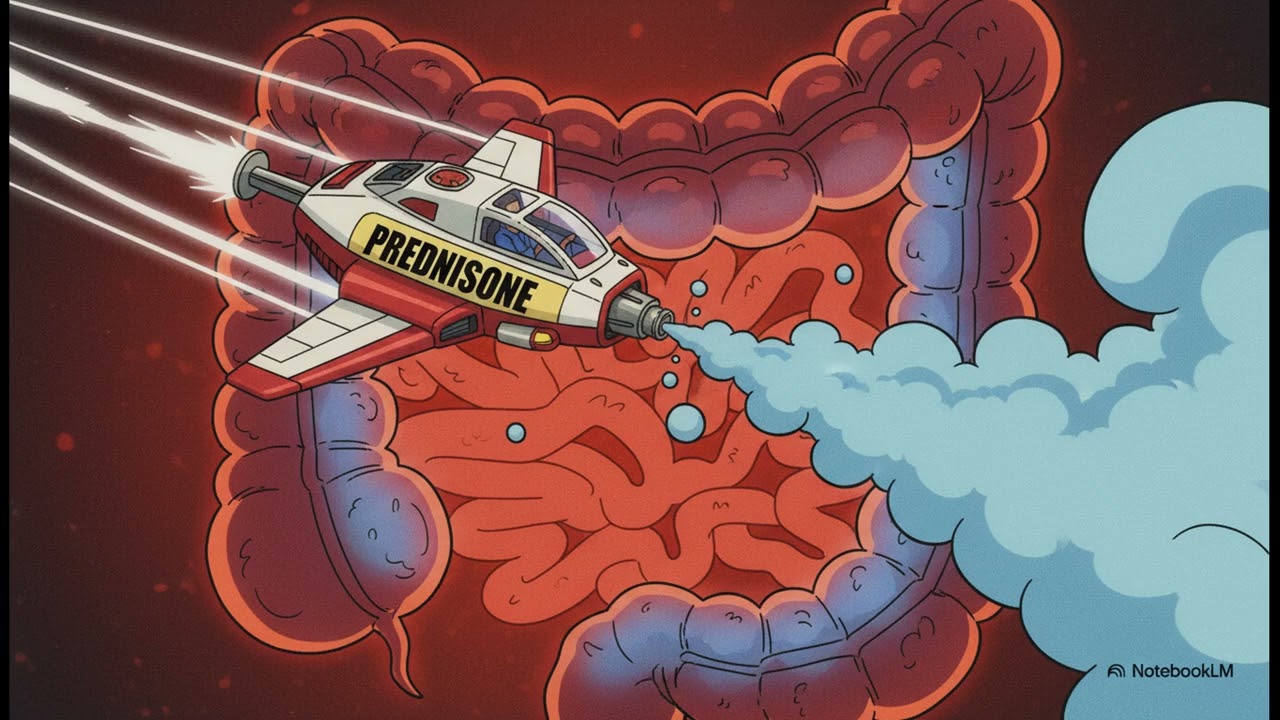 Crohn's Colitis: Explained Simply (A Beginner's Guide)This explainer defines Crohn's colitis as a type of Inflammatory Bowel Disease (IBD) where Crohn's disease is specifically located in the colon. Unlike ulcerative colitis, Crohn's inflammation affects the entire thickness of the bowel wall. It is an autoimmune condition where the body's security team mistakenly attacks healthy colon cells. The two main treatment goals are inducing remission (calming flare-ups immediately) and maintaining remission (long-term prevention of future flare-ups). Medications are categorized as a "toolkit": corticosteroids (fast-acting, short-term use), immunomodulators (long-term, steroid-sparing plan), and biologics (targeted, modern drugs for moderate to severe disease). Surgery is presented as an effective, strategic tool (not a sign of failure or last resort) used to manage complications like blockages or fistulas, or when medicines fail. Ongoing, proactive care ("treat-to-target") is necessary because even after surgery, inflammation returns in up to 90% of patients within three years if they are not on medical therapy.
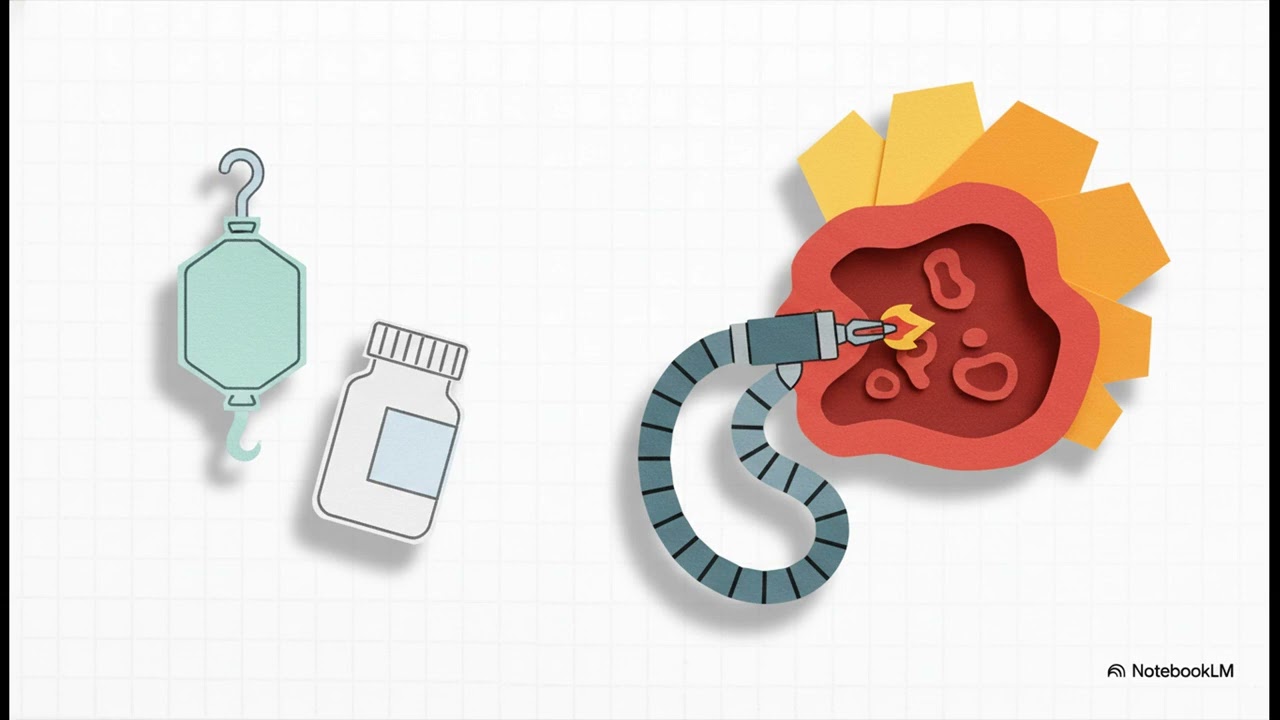 Radiation Proctitis: Dealing with Rectal Bleeding After Cancer TreatmentRadiation proctitis is inflammation of the rectum caused by radiation therapy. The content focuses on the chronic type, which appears 8 to 12 months after treatment and is caused by damage to small blood vessels. The main symptom is rectal bleeding (hemorrhagic proctitis), which results from the growth of new, fragile, spiderweb-like blood vessels called telangiectasias. Diagnosis involves reviewing radiation history and performing an endoscopy to visualize classic signs like easy fryability (tissue that bleeds easily). Highly effective treatments are generally non-surgical, focusing on endoscopic procedures. These include applying topical formalin (which seals the delicate vessels shut, with a success rate over 90%) and using Argon Beam Coagulation (ABC). Both methods work almost equally well. Surgery is considered a last resort only for specific, severe cases, due to the higher complication risk associated with operating on radiated tissue.
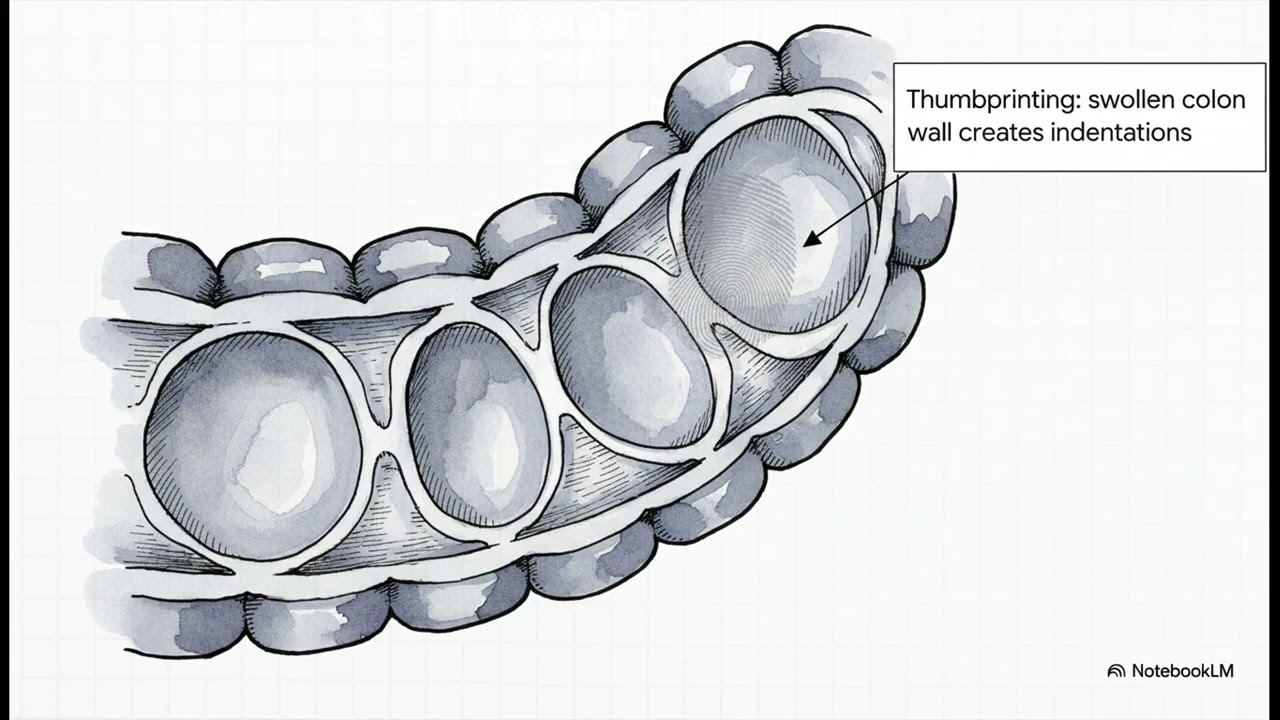 Ischemic Colitis: Understanding the Sudden Belly Pain and RecoveryIschemic colitis is caused by a temporary dip in blood flow to the small blood vessels of the colon, leading to inflammation and symptoms. It is critical to differentiate this temporary condition, which affects small vessels, from mesenteric ischemia, which involves major blood vessels and often requires emergency surgery. The condition frequently affects "watershed areas" in the colon—spots at the end of the blood supply line. Symptoms include sudden belly pain and bleeding. Diagnosis involves a physical exam, CT scan (which may show inflammation and "thumb printing"), and colonoscopy. For most people (over 80%), the condition is treatable with simple supportive care and no surgery. Supportive care includes bowel rest, IV fluids, and preventative antibiotics. The prognosis is very good, with a full recovery typically occurring within 1 to 2 weeks for the majority of patients.
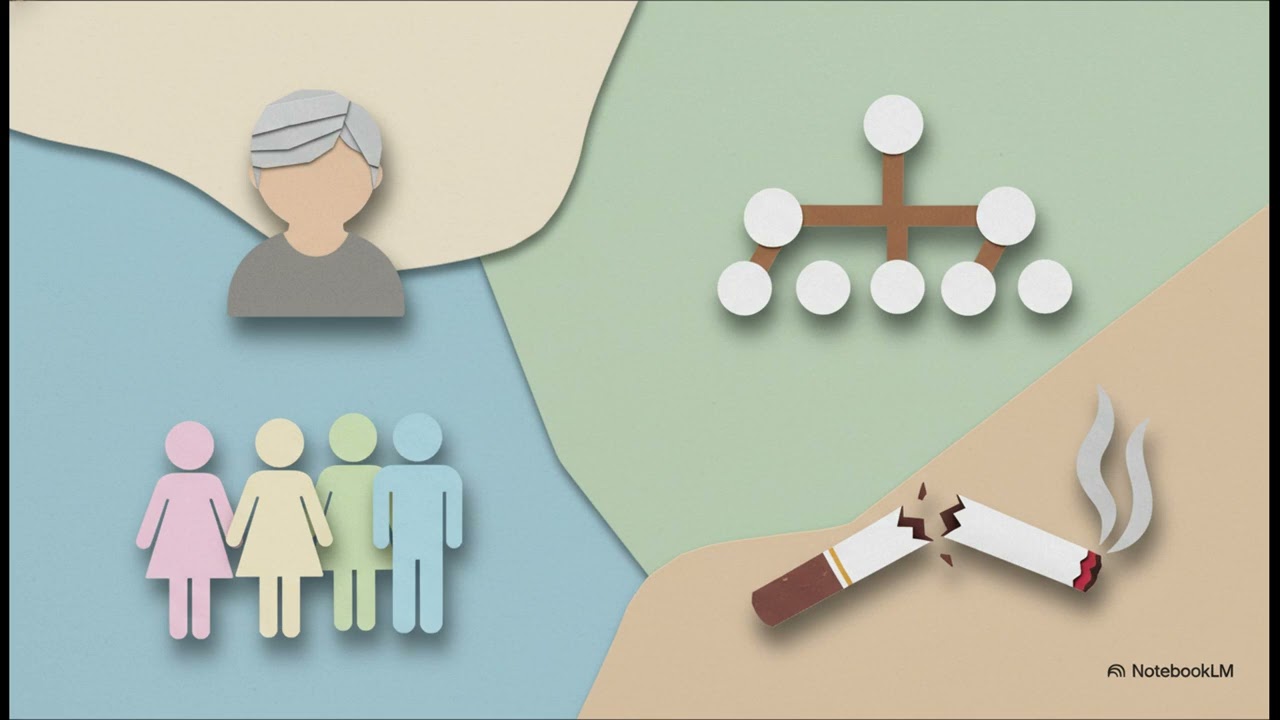 Stomach Trouble? Understanding Microscopic ColitisMicroscopic colitis (MC) is an inflammatory condition defined by chronic watery diarrhea, abdominal pain, and fatigue. Crucially, the inflammation is only visible under a microscope, meaning the colon looks totally normal during a colonoscopy. Definitive diagnosis requires a three-step process: symptoms, normal colonoscopy, and multiple biopsies (to account for patchy inflammation). The two subtypes are Collagenous Colitis (thick collagen band seen) and Lymphocytic Colitis (extra white blood cells seen). MC is more common in women, usually diagnosed around age 61, and risk is significantly increased by smoking. There is a strong link to other autoimmune disorders (40% of patients have another) and to common medications (NSAIDs, PPIs, SSRIs). The primary, highly effective treatment is the targeted corticosteroid Budesonide, which achieves remission in 68% of patients. MC is a benign chronic condition that does not increase the risk of colorectal cancer, and surgery is almost never needed.
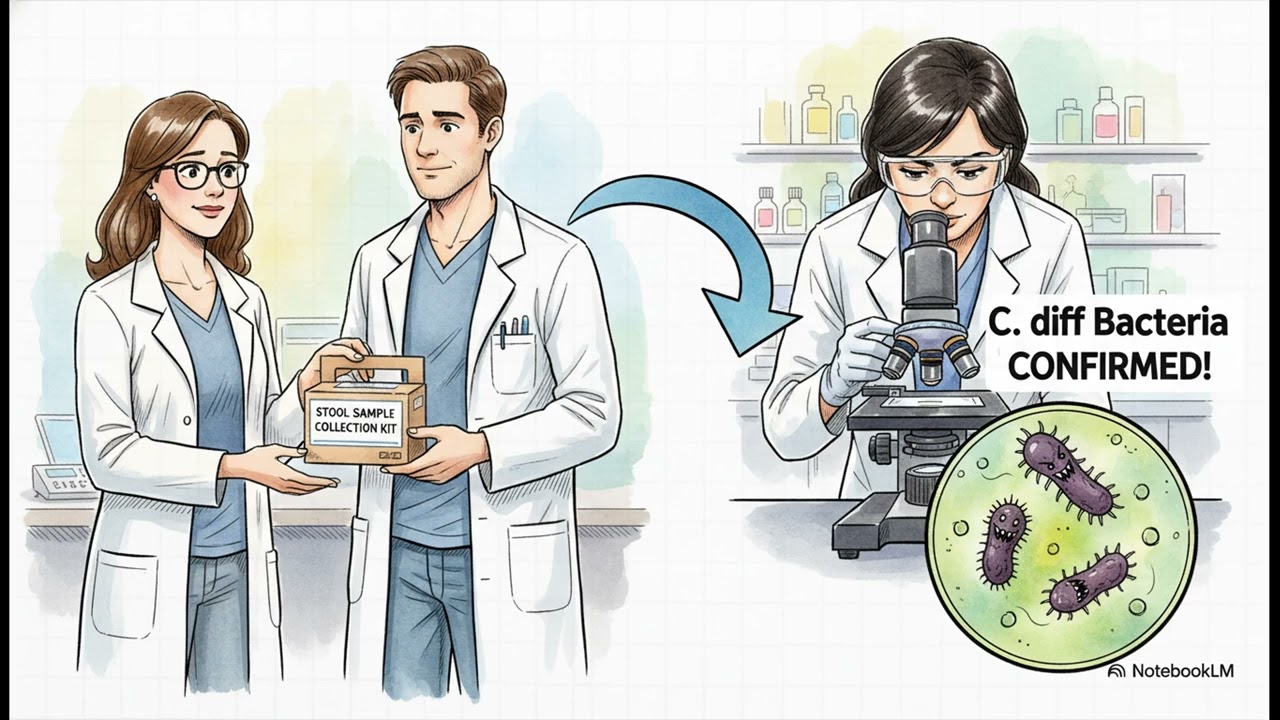 C Diff Infection: Why Antibiotics Can Make You Sick and How to Treat ItThis guide addresses C. difficile (C. diff) colitis, a condition often triggered by recent antibiotic use. C. diff is a bacterium that commonly lives in the gut but can rapidly multiply when antibiotics wipe out the protective good bacteria ("gut garden analogy"). As C. diff grows, it releases toxins that inflame the colon, causing symptoms like persistent watery diarrhea, abdominal cramping, and fever. Risk factors include recent antibiotic use, being 65 or older, and recent hospitalization. Diagnosis is simple, requiring a stool sample test for the bacteria or its toxins. Treatment for the infection involves targeted antibiotics, such as vancomycin. For frustrating recurrent C. diff, the highly effective treatment is a Fecal Microbiotic Transplant (FMT), which has cure rates consistently over 80%. Surgery to remove the colon is a very rare, last-resort option for life-threatening damage. Prevention emphasizes handwashing with soap and water and carefully discussing future antibiotic use with a doctor.
 Long-Term Constipation: Is It a Slow Highway or a Blocked Exit?This content explains chronic constipation (symptoms lasting over three months) and divides it into two distinct types using the "highway analogy". 1) Slow Transit Constipation ("Slow Highway") means the colon muscles lack the "oomph" to push waste along. 2) Functional Outlet Obstruction (or dyssynergic defecation, the "Blocked Exit Ramp") means the pelvic floor muscles tense up instead of relaxing at the end of the line. Diagnosis must identify the type. Slow Transit is diagnosed using a SITS marker study (counting tiny plastic rings swallowed by the patient on an X-ray five days later). Functional Outlet Obstruction is diagnosed using specialized tests like Anorectal Manometry or Defecography (a live-action X-ray video). Treatment is specifically tailored: Slow Highway gets laxatives and prescription medications to speed up the colon; Blocked Exit gets muscle retraining, most commonly through Pelvic Floor Physical Therapy using Biofeedback. Surgery is considered a last resort for severe, unresponsive cases.
 IBS Explained: Simple Guide to Irritable Bowel SyndromeThis resource provides a straightforward guide to Irritable Bowel Syndrome (IBS), a common condition defined by abdominal pain (at least one day a week) linked to changes in bowel movements, frequency, or consistency. It is emphasized that IBS is a benign condition; it does not physically damage the intestines or lead to diseases like cancer. IBS is broken down into subtypes (IBS-C, IBS-D, IBS-Mixed) based on stool consistency, often using the Bristol stool chart. Diagnosis is one of exclusion, where the doctor rules out other conditions (like celiac disease or infection) using blood tests, stool tests, or sometimes a colonoscopy. Treatment is highly personalized, starting with education, reassurance, and diet/lifestyle changes. Dietary focus includes soluble fiber (like psyllium) and the low FODMAP diet. Specific medications are chosen based on the patient's IBS subtype to target the main symptom. Having a strong, trusting relationship with the doctor is a massive part of successful management.
 Pelvic Bulges Explained: Rectum or Small Intestine?This resource addresses pelvic bulges (pelvic organ prolapse), which affect up to half of women who have had children. It focuses on two conditions: Rectocele (the rectum bulges forward into the vagina, a problem from the side) and Enterocele (the small intestine pushes down into the space, a type of internal hernia, a problem from above). Diagnosis relies on symptoms, as small rectoceles can be present in up to 93% of asymptomatic women. The gold standard test is Defecography, a specialized X-ray video that records the pelvic floor in action while the patient simulates a bowel movement to check the size of the bulge and if it traps stool. Surgery is recommended based on severe symptoms (e.g., needing to press on the vaginal wall to empty the bowels). Rectocele repair strengthens the weakened wall. Enterocele often requires a different procedure, such as a Sacrocolpopexy, to lift and secure the pelvic organs.
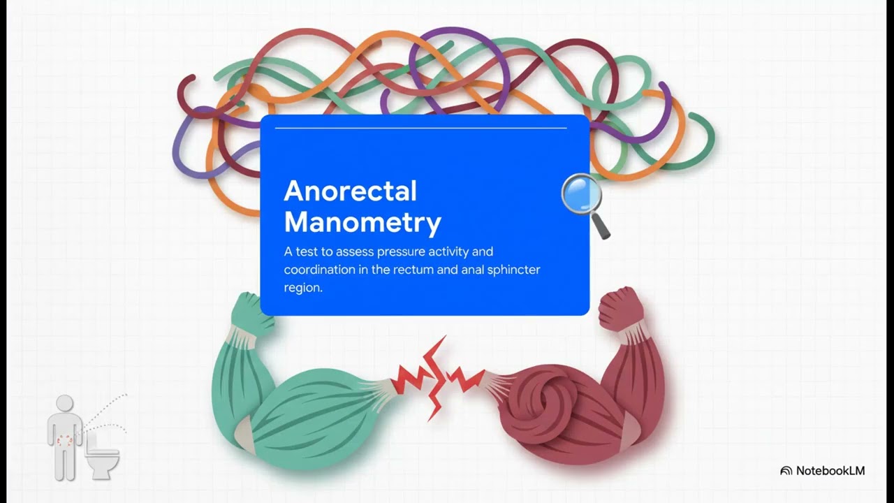 Dyssynergic Defecation: Why Can't I Empty My Bowels? (The Muscle Miscommunication)This content focuses on dyssynergic defecation, a common cause of chronic constipation (affecting up to 20% of people in North America). It results in a feeling of incomplete emptying or an "outlet obstruction". The core problem is a muscle miscommunication. When a person pushes to have a bowel movement, the pelvic floor muscles, which should relax, mistakenly tighten up, creating a blockage. Diagnosis starts with conversation and then involves specialized tests like Anorectal Manometry (to measure muscle pressures) and the Balloon Expulsion Test (to check for mechanical obstruction). Treatment is focused on retraining the body rather than stronger laxatives. The key treatment is specialized physical therapy using Biofeedback, which provides real-time information to help patients consciously learn to relax those pelvic floor muscles. This is a real, measurable condition that can be treated effectively, usually without surgery.
 Anal Warts (Condyloma Acuminata): HPV, Treatment, and PreventionThis video focuses on anal warts (condyloma acuminata), which are fleshy, non-cancerous growths. They are caused by the Human Papillomavirus (HPV), usually types 6 and 11. The virus is extremely common. Diagnosis typically starts with a visual exam, followed by an anoscopy to check for internal warts, and sometimes a biopsy. The goal of treatment is to remove all visible warts while preserving healthy skin. Treatment options include at-home medicated creams, in-office procedures (freezing/acid), or surgery. Crucially, treatment removes the growths but does not eliminate the underlying HPV virus, meaning recurrence is possible (4% to 29% recurrence rate). The HPV vaccine (Gardasil 9) is identified as the best strategy against HPV and HPV-related cancers. It also briefly addresses the rare and more aggressive variant, Giant Condyloma Acuminata (GCA), which requires wide surgical removal.
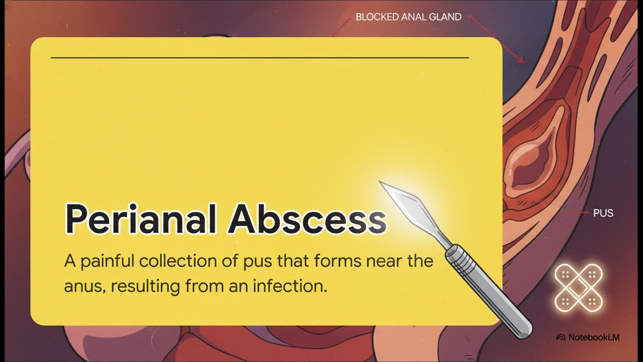 Perianal Abscess: Causes and the Power of DrainageA Perianal Abscess is a common, treatable pocket of pus that builds up near the anus. The cause in 90% of cases is a simple biological event: a tiny, normal anal gland gets blocked, trapping bacteria and leading to infection. Diagnosis is usually quick and straightforward based on the painful, swollen red bump visible during a physical exam. The definitive cure is immediate surgical drainage (a tiny cut to release the pus and pressure). The wound is left open to heal from the inside out. Post-procedure care involves sitz baths. Antibiotics are generally unnecessary for simple abscesses in healthy individuals, as drainage removes the source of infection. It is noted that about one-third of people who have an abscess drained may later develop an anal fistula (a persistent tunnel).
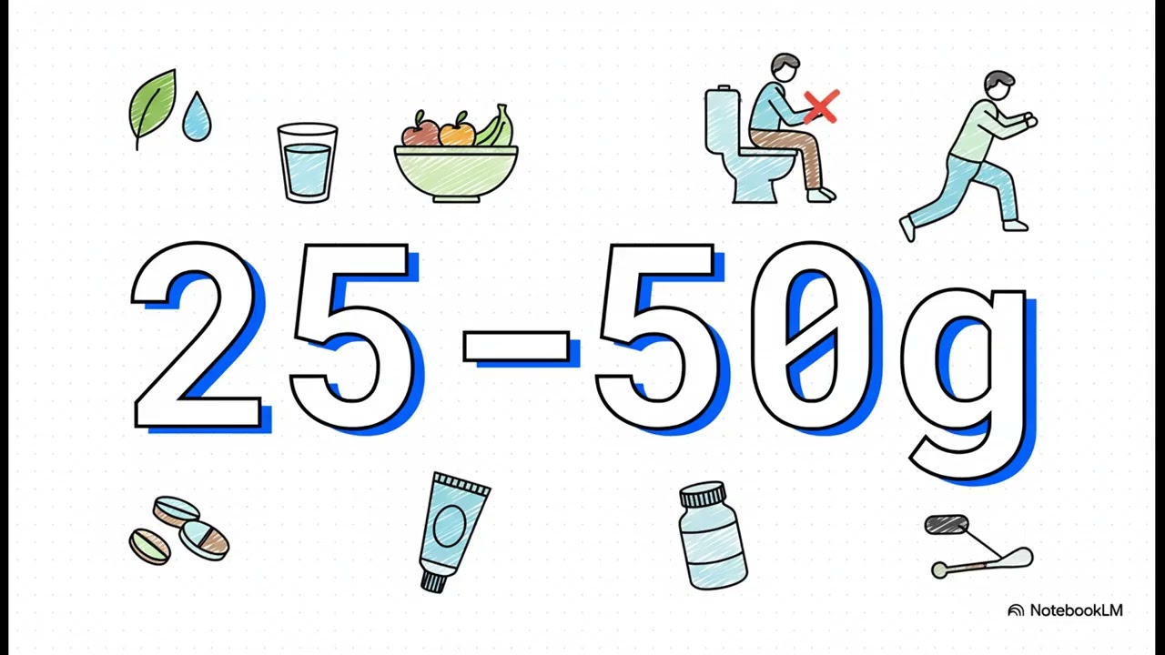 A Patient's Guide to HemorrhoidsThis video provides foundational information about hemorrhoids, explaining that they are normal parts of the anatomy (cushions of tissue and blood vessels) that aid in continence. Problems arise when increased pressure (due to straining, chronic constipation, or pregnancy) causes them to swell and cause symptoms. The two main types are internal (categorized by a grading system based on prolapse severity) and external hemorrhoids. Treatment begins with conservative care (the "most powerful line of defense"), focusing on reducing straining through increased fiber intake (25 to 50 g a day, often via supplements), hydration, proper toilet habits (avoiding long sitting times), and sitz baths. Medical options include quick office-based procedures like rubber band ligation (for less severe internal hemorrhoids) or surgery for more severe or stubborn cases, which is considered the gold standard effective fix.
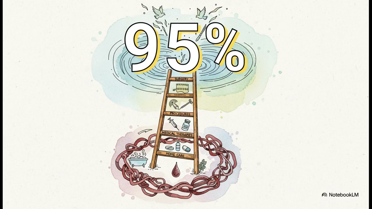 Anal Fissures Healing GuideAnal fissures are small cuts or tears in the sensitive skin of the anal canal, often causing sharp, tearing pain and bright red blood. They commonly occur in the back midline, which has naturally less blood flow. Fissures are categorized as acute (less than 6 weeks) or chronic (more than 6 weeks). The condition becomes chronic due to a vicious cycle where pain causes the internal anal sphincter muscle to spasm, cutting off blood flow needed for healing. First-line treatment involves conservative measures like increasing fiber/water intake, using stool softeners, and employing warm sitz baths to relax the spasm. Medical treatments are designed to break the cycle by relaxing the muscle; topical calcium channel blockers (like nifedipine cream) are considered the first-line medical treatment due to their effectiveness and fewer side effects compared to nitrates. For stubborn chronic fissures, the definitive surgical solution is a Lateral Internal Sphincterotomy (LIS), which is called the gold standard by the American Society of Colon and Rectal Surgeons and boasts success rates of 88 to 100%.
 Anal Fistula: How the Tunnel Forms and Surgical TreatmentAnal fistulas are often the second chapter of a process that begins with an anal abscess (a painful pocket of pus/infection). An abscess forms when a normal anal gland gets blocked. The fistula is a tunnel that remains open under the skin, connecting the blocked gland to the external drainage site. Fistulas are classified as simple (avoiding the main sphincter muscle) or complex (going through a large portion of the muscle), a distinction crucial for treatment planning. Mapping the fistula path, often using an Examination Under Anesthesia (EUA), is the first step. Treatment goals are infection control, preservation of bowel control, and permanent healing. Simple fistulas are typically treated with a fistulotomy (opening the tunnel). Complex fistulas require sphincter-sparing procedures (like the LIFT procedure) to close the tunnel while protecting the critical gatekeeper muscles.
 Anal Stenosis: Causes and Relief OptionsAnal Stenosis is the narrowing and tightening of the anal canal due to the replacement of flexible tissue with stiff scar tissue. The primary cause (90% of all cases) is a direct complication of a previous surgery, most commonly hemorrhoid surgery (hemorrhoidectomy). Other causes include deep anal fissures, Crohn’s disease, or radiation damage. The condition is classified as mild, moderate, or severe. Non-surgical treatment involves softening stool through diet changes and fiber supplements (bulking therapy) so that bowel movements act as a natural, gentle stretching tool. Gentle dilation (stretching with special tools) can be performed, but aggressive dilation must be avoided as it can worsen scarring. For moderate to severe cases, the surgical option is an anoplasty (flap surgery), which releases the scar tissue and reconstructs the area with a flap of healthy, flexible skin to widen the canal.