Intestinal Stomas
Dr. Allen Kamrava, MD, MBA, FACS, FASCRS
Episode Duration
36 MinutesPublished
May 19, 2026Category
Colorectal Surgery | Stoma Creation & Reversal | Ostomy Management | Surgical TechniqueHear Us on Spotify
Hear Us on Apple Music
Episode Summary
An intestinal stoma breaks the clean, binary endpoint most operations aim for: instead of resect, restore, and close, the surgeon builds an entirely new organ on the abdominal wall that the patient then manages for life. Working from McGee and Cataldo's chapter on intestinal stomas, this episode connects the intraoperative decisions to the lifelong management that follows — site selection, the orthogonal trephine, maturation, difficult-abdomen maneuvers, ischemia assessment, peristomal disease, high-output physiology, and the shifting evidence on when to reverse. It's aimed squarely at the board-testable, practice-altering detail.Key Highlights
- Why a stoma "shatters the binary" — you're constructing an artificial organ, not just ending an operation
- WOCN preoperative teaching: two visits cutting time-to-proficiency and length of stay, with a net cost saving per patient
- The ACS Ostomy Home Skills Kit for programs without in-house WOCN access
- Site selection beyond the classic stoma triangle — why a "perfect" supine mark fails when the patient stands, sits, or bears down
- The two-inch clear-skin perimeter, and why supra-umbilical placement beats infraumbilical in the morbidly obese
- Achieving a perfectly orthogonal trephine — and the gauze-sponge compression maneuver that keeps it perpendicular
- The ISI trial: full-thickness epidermal sutures cutting early peristomal excoriation versus classical intradermal fixation
- Stoma rods reconsidered — assignment bias in the 2019 meta-analysis, and the shift to three-to-five-day selective use
- Reach in a hostile abdomen: loop-end stoma, mesenteric pie-crusting, and the Prasad-type end loop
- ICG fluorescence versus the "eyeball test" for detecting early venous congestion
- The glass test-tube bedside exam that maps superficial versus sub-fascial ischemia
- Why a superficial-fascial colostomy can be observed but a flush ileostomy usually can't
- Peristomal pyoderma gangrenosum — pathergy, why biopsy misleads, and why debridement makes it worse
- Bleeding parastomal varices — why suture ligation fails ~85% of the time, and the role of TIPS
- The SGLT-1 physiology behind WHO oral rehydration solution — why the recipe needs sugar
- Anti-motility timing: why loperamide belongs 30 minutes before a meal, not after
- Food-bolus disimpaction — and the cardinal rule never to inflate the Foley balloon in the terminal ileum
- The Scandinavian EASY trial on early reversal — and the draconian selection criteria behind it
- Reversal during chemotherapy, the HASTA trial, and why stapled closures obstruct less than hand-sewn
- Purse-string skin closure as a dynamic drain, and protecting a urinary conduit's pedicle on reoperation
Who Should Listen?
This episode is valuable for:- Colon and rectal surgeons
- General surgeons who create and reverse stomas
- Surgical residents and fellows preparing for boards
- Wound, ostomy, and continence (WOCN) nurses
- Enterostomal therapy and ostomy care specialists
- Medical students on surgical rotations
- Anyone managing the lifelong care of ostomy patients
Key Topics Covered
| Topic | Discussion |
|---|---|
| Preoperative Preparation | WOCN teaching, site marking, and the two-inch clear-skin rule |
| Trephine & Maturation | Orthogonal path, compression maneuver, ISI-trial suture technique |
| Difficult Abdomens | Stoma rods, loop-end stoma, pie-crusting, Prasad-type end loop |
| Ischemia Assessment | ICG fluorescence and the bedside glass test-tube exam |
| Peristomal Disease | Pyoderma gangrenosum vs. appliance leak; bleeding varices |
| High-Output Stomas | SGLT-1 physiology, WHO ORS, anti-motility timing |
| Reversal | EASY trial timing, chemotherapy, HASTA closure technique |
| Closure & Crossover | Purse-string skin closure; protecting urologic conduits |
Featured Quote
"You take this highly vascular, microbiologically active mucosal surface and pull it through dynamic layers of muscle and fascia — you're essentially constructing an entirely new artificial organ right on the abdominal wall."— Colorectal Surgery Review, Episode Discussion
Transcript
Host:You know, usually when we evaluate a surgical intervention, the endpoints are beautifully binary — identify the pathology, achieve an R0 resection, restore continuity, and the operation is effectively complete. Guest:Right, it's very cut and dry. Host:But creating an intestinal stoma completely shatters that binary expectation. You're constructing an entirely new artificial organ on the abdominal wall — one the patient has to manage every day for the rest of their life… [0:06] Guest:Right, it's very cut and dry.
[0:07] Host:Exactly.
[0:07] Host:You identify the pathology, you achieve an R0 resection, you restore continuity and boom, the operation is effectively complete.
[0:15] Guest:Yeah, you pack up and go home.
[0:16] Host:But I mean, creating an intestinal stoma completely shatters that binary expectation.
[0:21] Guest:Oh, absolutely it does.
[0:22] Host:Because you take this highly vascular, microbiologically active mucosal surface and you pull it through dynamic layers of muscle and fascia, you're essentially constructing an entirely new artificial organ right on the abdominal wall.
[0:36] Guest:And it's an organ the patient has to manage every single day for the rest of their life.
[0:41] Host:Right.
[0:41] Host:So welcome to the deep dive.
[0:41] Host:This is our special colorectal surgery review series.
[0:41] Host:Glad to be here.
[0:41] Host:And just to set our mission for you, the listener today, we are strictly focusing on the clinically relevant board testable minutia.
[0:41] Host:We want the stuff that changes actual practice for the academic and practicing colorectal surgeon.
[1:01] Guest:The really high yield practice altering details.
[1:04] Host:Exactly.
[1:04] Host:We are unpacking the definitive text on intestinal stomus by McGee and Cataldo.
[1:10] Guest:It's a phenomenal resource.
[1:10] Guest:I mean truly comprehensive.
[1:14] Host:It really is.
[1:14] Host:We're going to examine the exact hemodynamics of why suture ligation for parastomal viruses fails so predictably.
[1:21] Guest:Yeah, that's a trap a lot of us fall into early on.
[1:23] Host:For sure, we'll also dissect the molecular Physiology of oral rehydration.
[1:23] Host:That makes our clinical advice sound, well, completely paradoxical.
[1:33] Guest:The high output stoma management is just fascinating and we're.
[1:36] Host:Going to look at some wild data on early stoma reversals that fundamentally challenges the rigid 812 week dogma we were all taught in residency.
[1:45] Guest:Right.
[1:45] Guest:The stuff that makes you rethink everything you learned.
[1:45] Guest:The beauty of this specific chapter is how it forces us to bridge the gap between the purely mechanical steps of indexedoma creation and then the incredibly complex lifelong medical management of the Ostoman.
[1:59] Host:Because they really are intrinsically linked, right?
[2:02] Guest:Absolutely.
[2:02] Guest:I mean, we are constantly searching for those small, highly specific intraoperative decisions that drastically alter a patient's postoperative trajectory.
[2:11] Host:Yeah, because it is very easy to treat the stoma as an afterthought.
[2:11] Host:You know at the end of a grueling 5 hour pelvic dissection, you're tired.
[2:18] Guest:Oh, you're exhausted, you just want to close.
[2:18] Guest:But the literature shows that surgical fatigue right there leads directly to debilitating complications later on.
[2:26] Host:It really does.
[2:26] Host:2:27WOCN Education and Optimal Stoma Site Selection
[2:26] Host:So let's start with the preoperative phase.
[2:30] Guest:Good place to start.
[2:31] Host:We all accept that preoperative education is beneficial, sure, but the text highlights Chowdhury's data regarding WOCN, the wound ostomy incontinence, nurses and their intervention.
[2:41] Guest:Yeah, the WOCN data is just incredible.
[2:43] Host:When we look at the actual clinical and financial metrics, how profound is the impact of catching these patients before the scalpel even hits the skin?
[2:51] Guest:The data is striking, especially for anyone tracking quality improvement or ER as protocols.
[2:51] Guest:I mean, Jowdry's perspective analysis demonstrated that just 245 minute preoperative visits with a WOCN.
[3:04] Host:Wait, just two visits.
[3:06] Guest:Just two and incorporating audio, visual aids and physical instruction, it decreased the time to post operative stoma proficiency from 9 days down to 5 1/2 days.
[3:15] Host:Wow, that's a massive drop in the learning curve.
[3:18] Guest:Exactly.
[3:18] Guest:And that accelerated independence directly reduced the hospital length of stay from 10 days down to 8.
[3:26] Host:Which obviously has huge financial implications for the hospital.
[3:29] Guest:Massive.
[3:29] Guest:When you model the reduction in unplanned provider encounters and emergency department visits for things like dehydration or appliance failure after discharge, it generated a net cost savings of over $2000 per patient.
[3:43] Host:$2000 just from 90 minutes of preoperative nursing time.
[3:47] Guest:Right, it is a staggering return on investment.
[3:50] Host:But what about smaller programs?
[3:50] Host:Like what if you're at a facility lacking robust in house WOCN access?
[3:56] Guest:Yeah, that's a real issue for those programs.
[3:56] Guest:The ACS Ostomy Home Skills Kit is a lifesaver.
[3:56] Guest:It provides an instructional model stoma allowing patients to develop muscle memory for pouching.
[4:07] Host:Which is huge, right?
[4:07] Host:Because they can practice before they are dealing with a swollen, highly sensate stoma postoperatively while loaded on pain meds.
[4:16] Guest:Precisely, you want them to.
[4:16] Guest:They have that mechanical practice beforehand.
[4:19] Host:Right.
[4:19] Host:OK.
[4:19] Host:So moving to the actual anatomical blueprinting, we frequently rely on the classic Stoma triangle, right?
[4:27] Guest:Yeah, the anterior superior iliac spine, pubic tubercles, umbilicus.
[4:31] Host:Right.
[4:31] Host:Or we try finding the intersection of the infrumbilical fat pad and the rectus sheath.
[4:38] Guest:The classic textbook landmarks.
[4:40] Host:But finding a mathematically perfect site on a supine anesthetized patient often completely fails when you see them in the clinic later.
[4:47] Guest:Oh, it fails all the time.
[4:48] Host:So what are the specific biomechanical and anatomical factors we have to account for dynamically?
[4:55] Guest:Well, The thing is, a site that appears perfectly planar when the patient is lying flat will often just disappear into a deep transverse skin fold the second they stand up.
[5:03] Host:Or it shifts directly onto their beltline the moment they sit up or bear down.
[5:07] Guest:Exactly the rigid criteria, and this is heavily tested on the boards by the way, is the necessity of a 2 inch perimeter of clear intact skin around the intact.
[5:17] Host:Two inches in every direction.
[5:19] Guest:Right, because that specific radius accommodates the adhesive flange of standard appliance systems without overlapping scars or the umbilicus or Bony prominences like the Asus you.
[5:30] Host:Really have to dynamically assess the abdominal wall shearing forces while the patient is awake.
[5:36] Guest:You have to standing, sitting, bending over.
[5:36] Guest:You have to see how the skin moves.
[5:40] Host:OK, let's unpack this.
[5:40] Host:If a patient has a massive panisha, does the stoma go below the fold where the the skin is tight or above it?
[5:48] Guest:Yeah, the infrumbilical approach in a morbidly obese patient is a classic trap.
[5:53] Host:Really because there's often a junior level instinct to place it there, perhaps thinking it provides a better cosmetic result by hiding the appliance.
[6:01] Guest:Right, but anatomically the abdominal wall is frequently much thinner above the umbilicus.
[6:06] Host:Oh interesting, so that helps with the pull through.
[6:08] Guest:Exactly.
[6:08] Guest:It significantly reduces the tension on the mesentery during the pull through and decreases the depth of your fascial trephine.
[6:15] Host:But what about functionally for the patient?
[6:17] Guest:From a purely functional standpoint, an infumbilical placement beneath a massive fold entirely eliminates the patient's line of sight.
[6:25] Host:Of course they literally can't see it.
[6:27] Guest:Right.
[6:27] Guest:If they cannot visually index the mucocutaneous junction, they cannot independently measure, cut and apply their wafer.
[6:27] Guest:It's impossible.
[6:36] Host:So they become dependent on someone else for basic hygiene.
[6:39] Guest:Exactly.
[6:39] Guest:So a super umbilical placement on the upper slope of the panis guarantees that essential visual access, which is the absolute cornerstone of independent stoma management.
[6:50] Host:That makes perfect sense.
[6:50] Host:6:51Orthogonal Trephine and ISI Trial Maturation Techniques
[6:50] Host:OK, so once we have the site dialed in perfectly, we move to the physical creation of the trephine in the OR.
[6:57] Guest:The fun part?
[6:58] Host:Right, and the text places immense emphasis on achieving a perfectly orthogonal path through the abdominal wall.
[7:05] Guest:It have to be a straight cylinder.
[7:06] Host:But if we are splitting the subcutaneous tissue and muscle, how do we prevent the layers from shearing or shifting once the retractors come out?
[7:14] Guest:Because that creates A tortuous pass for the bowel, right?
[7:17] Host:Exactly how do we avoid that?
[7:18] Guest:So the standard technique involves having your assistant wrap 2 fingers in a folded gauze sponge.
[7:23] Host:OK, inside the abdomen.
[7:25] Guest:Yes, they apply firm aggressive anterior pressure from inside the peritoneal cavity directly against the intended exit site.
[7:34] Host:So they're pushing up against the abdominal wall.
[7:36] Guest:Right.
[7:36] Guest:This maneuver physically compresses the tissue girth, essentially flattening the abdominal wall dynamically while you incise the fascial layers from the outside.
[7:45] Host:So you're cutting through compressed tissue?
[7:47] Guest:Exactly.
[7:47] Guest:By maintaining that compression, you ensure your Trephian cylinder remains perfectly perpendicular to the fascia.
[7:54] Host:Because if you veer obliquely.
[7:56] Guest:Or if the fascial layers slide against each other when the patient is extubated and the pneumoperitoneum is gone.
[8:02] Host:You essentially create a guillotine effect on the bowel.
[8:04] Guest:Exactly which leads directly to venous congestion or early mechanical obstruction.
[8:04] Guest:It's disastrous.
[8:09] Host:Which nobody wants.
[8:09] Host:So that brings us to maturation.
[8:09] Host:You're pulling the bowel out.
[8:09] Host:We're aiming for what, 2 to 3 centimeters of protrusion for odeostomies.
[8:18] Guest:Yeah, two to three for ileostomies and about 1 to 2cm for colostomies.
[8:21] Host:OK, so let's look at the ISI trial, because historically the brick technique was absolute dogma.
[8:27] Guest:Oh, it was gospel.
[8:27] Guest:Serumuscular bites to the dermis, rigidly avoiding the epidermis.
[8:33] Host:Right, because everyone was terrified of ectopic mucosal cells migrating and ruining the appliance seal.
[8:33] Host:But the ISI trial randomized patients to classical intradermal fixation versus full thickness epidermal sutures.
[8:45] Guest:Yeah, that trial really shook things.
[8:46] Host:Up What did we actually learn about the tissue dynamics there?
[8:49] Guest:The ISI trial data is a phenomenal example of surgical evolution.
[8:49] Guest:The full thickness epidermal sutures actually demonstrated a reduction in early postoperative peristomal excoriation.
[9:01] Host:A reduction, really.
[9:02] Guest:Yeah, dropping from 52% to 41% compared to the classical intradermal group.
[9:07] Host:That is a significant drop.
[9:07] Host:Why does it work better?
[9:10] Guest:The proposed mechanism is that full thickness sutures which incorporate that tough epidermal layer provide a vastly superior mechanical anchor.
[9:18] Host:Oh, because the dermis alone just doesn't have the same holding power.
[9:21] Guest:Exactly.
[9:21] Guest:It prevents micro retraction and stabilizes the averted mucosal spout against the shearing forces of the appliance and the abdominal wall.
[9:28] Host:What about the fear of epidermoid mucosal implantation creating those mucus secreting islands?
[9:34] Guest:It appears clinically insignificant, at least compared to the immediate benefit of having a mechanically robust leak proof ever version in that critical early healing phase.
[9:45] Host:Wow.
[9:45] Host:9:46Stoma Rods and Loop-End Stoma for Mesenteric Reach
[9:45] Host:OK, let's talk about mechanical stabilization for loop stonas.
[9:45] Host:The 2019 meta analysis on stoma rods is honestly fascinating.
[9:54] Guest:The very controversial paper.
[9:55] Host:It is because it concluded that rods do not prevent stoma retraction, but they do significantly increase the rates of stoma necrosis and peristomal dermatitis.
[10:04] Guest:Yeah, the complication rates were not great.
[10:06] Host:Based on that abstract alone, it sounds like we should abandoned them completely.
[10:06] Host:Burn all the stoma rods.
[10:06] Host:But the authors still advocate for conditional use.
[10:06] Host:How do we reconcile the meta analysis with the clinical reality of, say, a foreshortened mesentery?
[10:20] Guest:What's fascinating here is you have to critically evaluate the assignment bias in those.
[10:24] Host:Studies.
[10:24] Host:Oh, because surgeons aren't randomizing who gets a rod in real life.
[10:28] Guest:Exactly.
[10:28] Guest:Surgeons typically only deploy a rod in the most anatomically hostile cases.
[10:28] Guest:We're talking patients with profoundly thickened mesenteries, severe obesity or extreme posterior tension on the loop.
[10:41] Host:Right, so those specific stomas are already at a massive baseline risk for ischemia and retraction, regardless of whether you use ROD or not.
[10:49] Guest:Precisely.
[10:49] Guest:Furthermore, if you look at the methods, several protocols in that meta analysis left the rods in place for up to 14 days.
[10:57] Host:14 days.
[10:58] Guest:Yes, leaving a rigid plastic bridge compressing the peristomial skin for two weeks will inevitably cause pressure necrosis and dermatitis.
[11:06] Host:Well, yeah, obviously.
[11:06] Host:So how do we use them properly today?
[11:09] Guest:Modern practice limits rod placement to three to five days.
[11:09] Guest:It's just enough time for the initial centripetal tension to decrease as local edema subsides.
[11:17] Host:And allowing early fibrin adhesions to form at the fascial level.
[11:21] Guest:Exactly.
[11:21] Guest:So used selectively for a few days in a high tension scenario, it definitely remains A valid tool.
[11:26] Host:That makes total sense.
[11:26] Host:So when we encounter those anatomically hostile abdomens where the bowel simply will not reach the skin, like, say, an obese patient with severe mesenteric foreshortening, we need advanced maneuvers.
[11:41] Guest:Oh, you definitely need a backup plan.
[11:43] Host:The text details the Lupen Stoma pie crusting and the Prasad type end loop.
[11:43] Host:Walk us through the exact anatomical rationale for the loop end stoma when an end ileostomy simply won't reach.
[11:56] Guest:So the loop end stoma capitalizes on the vascular geometry of the terminal ileum.
[12:01] Host:OK, how?
[12:02] Guest:So frequently the absolute distal tip of your transective bowel is tightly tethered by the terminal branches of the ileocolic artery.
[12:09] Host:Right, it's pinned down.
[12:10] Guest:But the bowel segment, just five to 10 centimeters proximal to that tip, often has significantly more mobility due to the cascading arcade.
[12:17] Host:Oh, so instead of fighting that tethered tip.
[12:19] Guest:Exactly.
[12:19] Guest:You leave the close distal and intra abdominally and bring up the mobile proximal segment as a loop.
[12:25] Host:And you just mature it like a regular loop soma.
[12:27] Guest:You mature it identically to a standard loop.
[12:27] Guest:Soma, yes, but because the distal limb is completely blind enclosed, it functions purely as an instoma.
[12:36] Host:That is so clever it maximizes reach without requiring a dangerous central vascular high ligation.
[12:43] Guest:Exactly.
[12:43] Guest:12:43Pie-Crusting and Prasad Stoma for Difficult Abdomens
[12:43] Guest:It's a lifesaver.
[12:44] Host:And if we need to physically elongate the mesentery itself, the pie crusting technique is described.
[12:44] Host:But honestly, that sounds incredibly precarious given the venous plexus in there.
[12:54] Guest:It is extremely precarious.
[12:54] Guest:You need a steady hand.
[12:56] Host:What is the specific tissue layer we are targeting to gain that length without causing a massive hematoma?
[12:56] Host:So PIE.
[13:03] Guest:Crusting is essentially a micro surgical release of the visceral peritoneum.
[13:03] Guest:The limiting factor in mesenteric reach is rarely the primary vasorecta themselves.
[13:12] Host:Right, the vessels usually have some stretch.
[13:14] Guest:Exactly.
[13:14] Guest:The limiting factor is the restrictive peritoneal envelope encasing them.
[13:14] Guest:By making highly controlled sequential superficial incisions, scoring only the peritoneal surface exactly perpendicular to the axis of the blood vessels, you allow the underlying fibro fatty tissue to stretch.
[13:31] Host:And you can gain several crucial centimeters that way.
[13:34] Guest:You can, but, and this is a huge but, if your blade ventures even a millimeter too deep into the mesenteric fat, you will lacerate a primary vein.
[13:44] Host:And then you have a rapidly expanding hematoma that can compromise the entire conduit.
[13:49] Guest:Right.
[13:49] Guest:And then you're resecting more bowel.
[13:49] Guest:So caution is key.
[13:52] Host:Absolutely.
[13:52] Host:Now for the truly disastrous scenarios, let's say a perforated diverticulitis with massive fecal contamination, where a primary anastomosis is totally contraindicated.
[14:04] Guest:A true damage control situation.
[14:05] Host:Right.
[14:05] Host:And the bowel is so edematous it refuses to mature as a standard stoma.
[14:05] Host:We look at the Prasad type and loop.
[14:13] Guest:The Prasad stoma is an exceptional maneuver for those nightmare abdomens.
[14:17] Host:How does it work?
[14:18] Guest:You exteriorize both the proximal and distal bowel limbs through a single fascial trephine, just laying them side by side on the skin without maturing them into a classical unified loop.
[14:28] Host:You're not even suturing the mucosa to the skin yet.
[14:31] Guest:Not necessarily in the acute phase, no.
[14:31] Guest:You were simply isolating the pathology outside the abdominal cavity and securing it so it doesn't retract.
[14:38] Host:And what's the long term play there?
[14:39] Guest:The physiological advantage becomes incredibly apparent during the reversal.
[14:39] Guest:Because both limbs are already adjacent at the skin level, You can often perform a local stoma takedown and reanastomosis without ever re entering the hostel adhes peritoneal cavity.
[14:56] Host:Oh wow, you completely bypass the morbidity of a major reoperative laparotomy down the road.
[15:01] Guest:Exactly.
[15:01] Guest:It is brilliant forward thinking in a crisis.
[15:01] Guest:15:05ICG Fluorescence and Bedside Ischemia Assessment
[15:01] Guest:So assessing the profusion of all these maneuvers is obviously critical.
[15:01] Guest:Historically, we relied on capillary refill, mucosal color, you know, active bleeding from the cut edge.
[15:15] Guest:The classic eyeball test.
[15:16] Host:Right, but the text strongly advocates for ICG enhanced fluorescence.
[15:16] Host:Given that ICG requires specific equipment and timing, what are the specific failure points of traditional optical evaluation that make ICG superior for detecting estemia?
[15:31] Guest:Well, the truth is the human eye is notoriously poor and detecting early venous congestion.
[15:36] Host:Even for experienced surgeons.
[15:37] Guest:Even for the Masters, right, Because a Sonoma can appear perfectly pink and bleed briskly from its mucosal edge in the operating room simply because arterial inflow is intact.
[15:46] Host:Right, blood is getting in.
[15:47] Guest:But if the fascio trephine is slightly too tight, the low pressure venous flow is occluded.
[15:52] Host:And you won't see that immediately.
[15:54] Guest:No, that mucosal color won't shift to cyanotic or dusky until hours later in the recovery unit, well after you've closed the fascia and left the hospital.
[16:03] Host:Which is terrifying.
[16:03] Host:So how does ICG fix that?
[16:07] Guest:ICG fluorescence provides a real time objective map of the microvascular capillary bed.
[16:07] Guest:If the dye does not fluoresce brightly at the exact distal tip of the intended diversion, you have definitive proof of microvascular compromise right then and there.
[16:22] Host:Which prompts you to either resect further proximally or widen the fascial defect immediately.
[16:27] Guest:Exactly.
[16:27] Guest:It takes the guesswork out.
[16:29] Host:OK, let's follow that patient to the recovery ward.
[16:29] Host:Say it's post op day 2.
[16:29] Host:The nurse reports a dusky purple stoma.
[16:37] Guest:The call nobody wants to get.
[16:38] Host:Right.
[16:38] Host:We know it's ischemia, but the clinical decision tree hinges entirely on the depth of the necrosis.
[16:38] Host:How does the bedside test tube exam accurately map the ischemic demarcation line without taking the patient back to the OR?
[16:52] Guest:It's honestly an incredibly elegant piece of bedside diagnostics.
[16:55] Host:Walk us through it.
[16:56] Guest:You take a standard lubricated clear glass laboratory test tube and gently advance it into the stoma lumen.
[17:02] Host:OK, a glass test tube.
[17:04] Guest:Right, And by shining a pen light directly down the center of the glass, the tube acts as a 360° speculum.
[17:04] Guest:You are visually tracing the mucosal viability deep into the abdominal wall.
[17:17] Host:That's brilliant.
[17:17] Host:So what are you looking for?
[17:19] Guest:If the mucosa transitions from black to healthy pink above the level of the anterior rectus fascia, you have super fascial ischemia.
[17:27] Host:But if the necrosis extends deeper.
[17:29] Guest:If it extends down through the fascial layer into the peritoneal cavity, you have sub fascial ischemia.
[17:35] Host:And the management diverges sharply there.
[17:35] Host:Sub fascial is obviously a mandatory trip to the OR for immediate revision to event free perforation and fecal peritonitis.
[17:44] Guest:You have to go back, no question.
[17:46] Host:But what about superfascial ischemia?
[17:46] Host:Because the text draws a hard line between observing A superfascial colostomy versus revising A superfascial ileostomy.
[17:46] Host:What drives that specific divergent?
[17:58] Guest:It all comes down to the physical properties of the effluent and dermatological physics.
[18:01] Host:OK, break that down.
[18:01] Host:Let's start with the colostomy.
[18:04] Guest:If a colostomy suffers superfascial sloughing, the necrotic tip will demarcate and eventually fall off.
[18:04] Guest:This leaves a flush or even slightly retracted stoma at the skin level, right?
[18:04] Guest:Because colonic output is generally formed in solid, a skilled WOCN can often manage a flush colostomy with convex appliances and paste.
[18:04] Guest:The patient can live with it safely.
[18:26] Host:But small bowel effluent is totally.
[18:27] Guest:Different oh completely small bowel effluent is liquid and loaded with highly active proteolytic and lipolytic enzymes.
[18:34] Host:So a flush ileostomy is basically pouring acid on the skin.
[18:38] Guest:Exactly, it will continuously direct caustic liquid, laterally undermining the appliance seal and rapidly dissolving the epidermis.
[18:38] Guest:A flush ileostomy is a dermatological catastrophe.
[18:49] Host:So early operative revision is almost always favored in a physiologically fit patient with a flush ileostomy.
[18:55] Guest:Yes, you really don't want to try and manage that conservatively if you don't have to.
[18:55] Guest:18:58Differentiating and Treating Peristomal Pyoderma Gangrenosum
[18:55] Guest:Let's dive deeper into parastomal dermatology, specifically the differentiation between a severe neglected alliance leak and pyoderma gangrenosum.
[19:07] Guest:PPG a nightmare to treat.
[19:09] Host:Because the pathogen mechanism in PPG is fascinating.
[19:09] Host:If a resident sees a deep purulent ulcer and aggressively debrides it, it literally explodes.
[19:19] Guest:Yeah, surgical trauma feeds it.
[19:20] Host:Why do standard tissue biopsies fail to give us a definitive diagnosis here, and what specific morphological borders are we looking for clinically?
[19:29] Guest:So Peter Mcgangrenosum is an immune mediated inflammatory ulceration heavily associated with IBD.
[19:29] Guest:The hallmark as you mentioned is pathogy, which is a massive dysregulated neutrophilic response triggered by minor physical trauma.
[19:29] Guest:So.
[19:44] Host:Scalpel blade essentially pours gasoline on the fire.
[19:47] Guest:Exactly.
[19:47] Guest:And biopsies are notoriously unhelpful because they merely demonstrate a dense, sterile, neutrophilic infiltrate.
[19:54] Host:Which looks exactly like what?
[19:55] Guest:Which is entirely indistinguishable from a severe contact dermatitis or a secondary bacterial infection under the microscope.
[20:02] Host:Wow, so you really must rely on the clinical evolution.
[20:05] Guest:You have to think about it.
[20:05] Guest:Chemical excoriation from an appliance leak works from the outside in.
[20:05] Guest:It creates a weeping superficial erosion with flat raw borders that perfectly match the pooling pattern of the effluent.
[20:17] Host:Right.
[20:17] Host:It looks like a chemical burn under the wafer.
[20:19] Guest:Exactly.
[20:19] Guest:But PPG works from the inside out.
[20:19] Guest:It begins as an intensely painful hemorrhagic pustule or nodule that rapidly cavitates.
[20:29] Host:And the borders.
[20:29] Guest:The borders are the key.
[20:29] Guest:They are raised highly violaceous or purplish in color and physically undermined, meaning you could actually slip a probe underneath the edge of the surrounding intact skin.
[20:41] Host:Oh wow.
[20:41] Host:That's a very distinct clinical picture.
[20:41] Host:And the treatment requires A systemic approach, right?
[20:47] Guest:Yeah, starting with intralesional steroids, escalating to topical tacrolimus, and eventually systemic biologics like infliximab.
[20:55] Host:Yet the text notes that even with optimal medical management, over 50% of these patients require surgical residing.
[21:02] Guest:Which carries a massive recurrence risk at the new site.
[21:05] Host:It perfectly illustrates that the stoma is not just an isolated mechanical pipe, you know.
[21:05] Host:It is a vascularized mucosal surface highly susceptible to systemic autoimmune pathways.
[21:14] Guest:Could not have said it better.
[21:14] Guest:21:15Hemodynamic Management of Bleeding Parastomal Varices
[21:14] Guest:Speaking of systemic pathways, let's examine parastomal viruses.
[21:19] Guest:Another complex issue.
[21:21] Host:Very much so in patients with portal hypertension, particularly PSC associated IDD, we see this port of systemic shunting.
[21:21] Host:The high pressure portal blood in the stomach mesentery seeks the low pressure systemic venous circulation of the abdominal wall.
[21:37] Guest:Creating a caput medusae right at the miko cutaneous junction.
[21:40] Host:Exactly, And when these bleed it can be terrifying.
[21:40] Host:Why does simple suture ligation fail so predictably here with an 85% failure rate?
[21:50] Guest:Suture ligation fails because it address the symptom while completely ignoring the hemodynamics.
[21:54] Host:Because the pressure is still there.
[21:56] Guest:Right, you are dealing with friable hyperkeratotic varicosity subjected to massive portal pressures.
[21:56] Guest:If you throw a figure of eight stitch and like it a bleeding vessel, the portal pressure doesn't decrease at all.
[22:08] Host:It just finds a new path.
[22:09] Guest:Exactly.
[22:09] Guest:It simply forces the blood into an adjacent paper thin collateral vessel millimeters away, which immediately ruptures under the new pressure load.
[22:09] Guest:Is a feudal game of hemodynamic whack a mole?
[22:20] Host:So the gold standard is portal decompression via TPS, which boasts an 80% durable cure rate.
[22:26] Guest:TPS is excellent if they can tolerate it.
[22:28] Host:But for the patient who is not a TPS candidate, perhaps due to severe right heart failure or advanced encephalopathy, we have to consider mucarcutaneous disconnection.
[22:38] Guest:A very challenging operation.
[22:39] Host:Walk us through the specific anatomy of that salvage procedure.
[22:44] Guest:Eucocutaneous disconnection is a highly morbid, technically demanding operation.
[22:44] Guest:You are making a circumferential incision exactly at the mucocutaneous junction, dissecting the bowel away from the skin and subcutaneous tissue down to the fascial level.
[22:58] Host:So you're freeing the stoma completely?
[23:00] Guest:Yes, you are essentially dismantling the entire portisystemic collateral network by force.
[23:00] Guest:It involves meticulously identifying and ligating dozens of massively engorged, thin walled venous trunks.
[23:13] Host:I imagine the blood loss can be profound.
[23:15] Guest:It can be terrifyingly fast.
[23:15] Guest:Once disconnected, you remature the stoma to fresh skin.
[23:20] Host:But it's not a permanent cure, right?
[23:21] Guest:No, it is purely A temporizing measure as the portal hypertension will eventually Dr. new viruses to form, but it can absolutely salvage a patient who is actively exsanguinating.
[23:31] Host:Let's pivot to high output stomas, generally defined as over 1200 to 1500 milliliters a day, which can precipitate rapid acute kidney injury.
[23:31] Host:23:40SGLT1 Mechanics of WHO Oral Rehydration Solution
[23:31] Host:We see this so often in new ileostomies.
[23:42] Host:The management algorithm is incredibly strict.
[23:42] Host:The first line is fluid restriction combined with The Who oral rehydration solution, and the text provides the exact milligram recipe. 38 teaspoon salt 14 teaspoon salt Substitute 1.5 teaspoons baking soda and over 2 tablespoons of sugar in a liter of water.
[24:02] Guest:It's very specific.
[24:03] Host:Here's where it gets really interesting.
[24:03] Host:We tell these patients to avoid concentrated sweets to prevent osmotic diarrhea.
[24:03] Host:But The Who or S recipe explicitly calls for 2 tablespoons of sugar.
[24:03] Host:How does that make sense?
[24:15] Guest:This is arguably one of the most critical physiological concepts in stoma management.
[24:15] Guest:The intestinal absorption of water in the small bowel is not purely passive.
[24:15] Guest:It relies heavily on the sodium glucose linked transporter, or SGLT 1, located on the apical membrane of the interocyte.
[24:31] Host:OK, so it's an active transport mechanism?
[24:33] Guest:Right, SGLT 1 is a supporter that operates on a rigid stoichiometric ratio.
[24:33] Guest:It requires the simultaneous binding of exactly 1 molecule of glucose and one molecule of sodium to induce the conformational change that transports both into the cell.
[24:48] Host:They have to go together.
[24:48] Guest:Exactly.
[24:48] Guest:And once they're inside the cell, water passively follows this massive osmotic gradient into the bloodstream.
[24:55] Host:So what happens if they just drink a regular sugary drink?
[24:58] Guest:If a patient drinks a soda, they're flooding the lumen with glucose but providing almost zero sodium.
[24:58] Guest:The SGLT 1 symporters cannot function without the sodium pair.
[25:07] Host:So the glucose is just left behind.
[25:08] Guest:You remain stranded in the bowel lumen, and because it's an act of osmolite, it exerts a massive osmotic pull, dragging water out of the mucosal cells and creating profound osmotic diarrhea.
[25:19] Host:That makes perfect sense, but the WHOORS avoids this.
[25:23] Guest:The Who or S is mathematically calibrated.
[25:23] Guest:It provides the exact optimal molar ratio of sodium to glucose.
[25:23] Guest:Every single glucose molecule is perfectly paired with the sodium ion, hyperactivating those SGLT 1 transporters.
[25:37] Host:So they rapidly pump the ions into the enterocyte and the water just follows right behind.
[25:41] Guest:Exactly, the sucrose isn't there for caloric nutrition at all.
[25:41] Guest:It is literally the biochemical catalyst required to activate the sodium pump.
[25:49] Host:Wow, that perfectly explains the strict recipe.
[25:49] Host:25:53Anti-Motility Agents and Food Bolus Obstruction
[25:49] Host:Now the second step in the algorithm is anti motility agents like loparamide or diphenoxylate atropine.
[25:49] Host:But the timing is everything.
[26:01] Guest:Timing is crucial.
[26:02] Host:Taking it postprandially is useless.
[26:02] Host:Why must it be taken exactly 30 minutes prior to the meal?
[26:08] Guest:Well, the goal of an anti motility agent is to increase the mucosal contact time of the chyme.
[26:08] Guest:If you wait until after the meal is ingested, the gastrocolic reflex has already initiated massive peristaltic waves.
[26:20] Host:And the chyme is rapidly transiting the shortened vowel before the drug even kicks in.
[26:24] Guest:Exactly.
[26:24] Guest:Administering the medication 30 minutes prior preemptively paralyzes the smooth muscle of the gut.
[26:30] Host:So when the food gets there.
[26:32] Guest:It ensures that when the food bolus arrives, it sits in the absorptive segments of the jejunum and ileum long enough for those SGLT 1 transporters to actually extract the fluid.
[26:41] Host:Right and then steps three and four are anti secretory therapies like PPI's and eventually things like opium tincture if needed.
[26:48] Guest:Yes, escalating is necessary.
[26:50] Host:Let's briefly look at a mechanical issue, the food bolus obstruction.
[26:50] Host:You know, poorly chewed popcorn, mushrooms or nuts physically impinging the stoma right at the fascial level.
[27:01] Guest:Very common ER presentation.
[27:03] Host:The bedside disinfection technique involves gently passing a 14 to 18 French Foley catheter just below the fascia and irrigating with 30 to 50 mils of water.
[27:03] Host:But the absolute cardinal rule is to never ever inflate the Foley balloon.
[27:03] Host:What is the specific tissue architecture of the terminal ileum that makes balloon inflation so incredibly dangerous here?
[27:26] Guest:Unlike the robust, thick muscularis propria of the rectum that we are used to dealing with, the wall of the terminal ileum is remarkably thin.
[27:34] Host:Especially when it's adematous.
[27:36] Guest:Exactly.
[27:36] Guest:When it's adematous and stretched over a tight fascial defect, it's like wet tissue paper.
[27:36] Guest:The radial force exerted by inflating a standard Foley balloon will easily exceed the tensile strength of the eyleal serosa and muscularis.
[27:48] Host:Leading to an immediate iatrogenic transmural perforation.
[27:53] Guest:Yes, you instantly turn a simple mechanical impaction into a catastrophic intraperitoneal fecal spell.
[27:59] Host:Just a total disaster.
[28:00] Guest:So you must rely entirely on the gentle hydro dissection of the water flush, never mechanical balloon dilation.
[28:07] Host:Such an important warning.
[28:07] Host:28:08Early Reversal, Chemotherapy, and Anastomosis Techniques
[28:07] Host:OK, we have navigated the creation and the complications.
[28:07] Host:Let's look at the end game stoma reversals.
[28:14] Guest:The light at the end of the tunnel for these patients.
[28:16] Host:Truly preoperative objective evaluation like contrast studies or endoscopy to ensure distal and asthmatic patency and rule out leaks or strictures is non negotiable.
[28:27] Guest:Oh, mandatory.
[28:28] Host:But the real controversy lies in the timing.
[28:28] Host:The H 12 week dogma was deeply ingrained in our training to allow pelvic adhesions to soften.
[28:28] Host:Yet the Scandinavian Easy trial investigated closing loop ileostomy is at just 8 to 13 days post.
[28:43] Guest:OP yeah, the early closure data is provocative.
[28:45] Host:How do we justify operating during the exact window when adhesions are supposedly at their most hostile and hypervascular?
[28:52] Guest:Well, the theory behind early closure is that at post op day 8, the fibrinous adhesions haven't yet organized into those dense fibrous of vascular bands we see at week four or five.
[29:03] Host:So they're still kind of soft.
[29:05] Guest:Right, the tissue planes can often still be bluntly separated.
[29:05] Guest:The trial demonstrated that in highly selected patients, early closure was statistically as safe as waiting 12 weeks.
[29:15] Host:And it spared the months of having a stomach.
[29:18] Guest:Exactly.
[29:18] Guest:It spared patients three months of dehydration risk, appliance leaks and skin excoriation.
[29:24] Host:But if the easy trial shows early closure is safe, why isn't every surgeon doing this at day 8?
[29:24] Host:Is it just surgical inertia or is the risk profile actually higher than the trial suggests for the average patient?
[29:36] Guest:If we connect this to the bigger picture, you have to look at the inclusion criteria.
[29:36] Guest:The patient selection criteria in that trial were absolutely draconian.
[29:44] Host:Oh, really?
[29:45] Guest:Yes, nearly 70% of screened patients were completely excluded from the study.
[29:49] Host:70%.
[29:50] Guest:If a patient had even a hint of a micro leak, slight tachycardia, or any systemic inflammatory response, they were disqualified.
[29:50] Guest:The clinical calculus is brutal.
[29:50] Guest:A leak following a standard 12 week reversal is usually a localized pelvic collection.
[30:05] Host:Right.
[30:05] Host:Manageable.
[30:06] Guest:But a leak following an 8 day reversal in a patient still recovering from the massive systemic hit of a low anterior resection is a lethal intra abdominal disaster.
[30:16] Host:So early reversal is technically feasible, but patient selection is paramount.
[30:16] Host:They have to be recovering Florida State.
[30:22] Guest:Absolutely, Florida State.
[30:23] Host:We also see shifting data regarding reversals during adjuvant chemotherapy.
[30:23] Host:The historical fear was that systemic chemotherapy completely halts collagen deposition and wound healing.
[30:33] Guest:Mandating we leave the stoma until the entire treatment course is completed.
[30:37] Host:Right.
[30:37] Host:What is the emerging literature suggesting about the actual cellular impact of chemo on the anastomotic staple line?
[30:45] Guest:We are actually learning that the inhibitory effect of modern fluorosil based or oxaliplatin based regimens on late stage collagen cross linking might be clinically overstated regarding bowel healing.
[30:57] Host:Really.
[30:57] Host:So the bowel it all heals anyway.
[30:59] Guest:Retrospective data increasingly shows that performing a loop closure mid chemotherapy cycle has comparable anastomotic leak rates to waiting until completion.
[31:08] Host:That's a huge quality of life win for the patient.
[31:11] Guest:It is the bowel just possesses an incredible intrinsic regenerative capacity that seems somewhat protected from the generalized macrophage inhibition caused by chemo.
[31:11] Guest:This allows us to potentially restore continuity much earlier in the oncologic pathway, though obviously it requires meticulous multidisciplinary coordination with medical oncology.
[31:31] Host:Absolutely.
[31:31] Host:Regarding the surgical technique for reversal, let's talk about the HASTA trial.
[31:31] Host:It compared hand sewn closures to stapled functional end to end anastomosis.
[31:40] Guest:A great trial.
[31:41] Host:The leak rates were equivalent, so the ASCRS guidelines accept both, but the stapled closures resulted in significantly fewer early post operative bowel obstructions.
[31:41] Host:What is the exact geometric and physiological reason a hand sewn closure obstructs more frequently?
[31:57] Guest:It comes down to tissue edema and luminal geometry.
[31:57] Guest:A transverse hand sewn closure of loop stoma inherently folds the tissue.
[31:57] Guest:This creates A relatively narrow fixed luminal diameter right at the suture line in the initial 72 hours postoperatively.
[31:57] Guest:Inflammatory tissue edema is inevitable.
[32:15] Host:And that edema swells inward.
[32:17] Guest:Exactly.
[32:17] Guest:It rapidly encroaches on that narrow hand sewn lumen, causing a functional early obstruction.
[32:23] Host:But the stapler avoids this.
[32:25] Guest:A stapled side to side functional end to end anastomosis creates a massively wide overlapping common channel.
[32:25] Guest:Even when Maxwell tissue edema peaks, the cross-sectional area of that stapled lumen is so large that it easily maintains patency for chyme to pass.
[32:42] Host:It just has more runway.
[32:43] Guest:Exactly.
[32:44] Host:Finally, we close the skin.
[32:44] Host:32:46Purse String Closure and Urologic Conduit Protection
[32:44] Host:The text is definitive regarding purse string skin closure over linear closure.
[32:44] Host:When we close linearly, we are trapping a highly contaminated subcutaneous space.
[32:55] Guest:The worst thing you could do for an infection risk.
[32:57] Host:How does the purse string physically alter those infection dynamics?
[33:00] Guest:Well, a linear closure completely seals the dermis over an inverted cone of contaminated subcutaneous fat.
[33:00] Guest:You're creating the perfect anaerobic incubator for a surgical site infection.
[33:10] Host:Right, it's going to get infected.
[33:11] Guest:Almost always, but a purse string closure, which is a subcuticular stitch drawn tight but leaving a small central three 5mm gap, acts as a continuous dynamic drain.
[33:23] Host:Oh, so it just lets everything out?
[33:25] Guest:It allows serosanguinous fluid and liquefying fat to egress freely, drastically dropping the wound infection rate.
[33:25] Guest:And honestly, it eventually contracts into a highly cosmetic little dimpled scar that patients are quite happy with.
[33:39] Host:That makes total sense.
[33:39] Host:Before we conclude there is a critical urologic crossover that frequently appears on the boards.
[33:39] Host:As colorectal surgeons, we often operate in abdomens containing genitorinary diversions like an Elio conduit, Indiana Pouch.
[33:39] Host:Mitrofanov.
[33:53] Guest:Right, we share the pelvis.
[33:54] Host:The urologist has isolated a segment of terminal ileum, attached the ureters, and restored bowel continuity with a new ileostomy.
[33:54] Host:When we are back in that abdomen dissecting adhesions, what is the exact vascular orientation we must protect to avoid de vascularizing that urologic conduit?
[34:12] Guest:This is a classic high stace anatomical trap.
[34:12] Guest:The blood supply to that isolated urinary conduit, it's dedicated mesenteric pedicle, is located anatomically inferior to the newly constructed ileostomy.
[34:26] Host:Inferior.
[34:27] Guest:The conduits mesentery dives deep into the pelvis toward the retroperitoneum.
[34:27] Guest:If you are blindly sweeping or firing an energy device to clear adhesions around that bowel and astomosis, and you inadvertently transect that inferior pedicle, you instantly infarct the entire urinary diversion.
[34:27] Guest:You have just triggered A catastrophic urologic emergency.
[34:27] Guest:You must explicitly identify and preserve that dependent mesentery before touching the bowel.
[34:55] Host:That is the exact level of anatomical vigilance we need.
[34:55] Host:34:58Balancing Technology and Physiological Limits in Stoma Care
[34:55] Host:Well, we've navigated the preoperative blueprint, the execution in the hostile abdomen, the molecular Physiology of output management, and the shifting paradigms of reversal.
[35:07] Guest:It's been a massive review.
[35:09] Host:It really has, but I want to leave everyone, you the listener, with a final thought to consider.
[35:09] Host:We spent our careers mastering the perfect auversion, preserving the vascular pedicles and mitigating these profound complications.
[35:09] Host:But look at the trajectory of our field.
[35:09] Host:It's changing fat as advanced biological therapies achieve deeper and deeper mucosal healing in IBD, and as our robotic platforms allow us to confidently construct anastomosis just millimeters from the dentate line, ones that would have definitely been permanent colostomies A decade ago.
[35:09] Host:Will the permanent end stoma eventually become a historical surgical relic?
[35:09] Host:Or as our patient population becomes increasingly older, frailer, and less capable of surviving the systemic hit of a pelvic leak, will the perfectly crafted brick alleostomy remain one of the most vital life saving maneuvers in a colorectal surgeons hands?
[35:57] Guest:It is the defining tension of our specialty, you know, balancing aggressive technological preservation with a fundamental physiological limits of human healing.
[35:57] Guest:It's a great question.
[36:06] Host:And mastering that balance requires knowing the.
[36:06] Host:Thank you for joining us on this colorectal surgery review, and we will see you on the next deep dive.
Related Episodes
Abdominal Wall Reconstruction and Parastomal Hernia
Radiation, Microscopic, and Ischemic Colitis
Clostridium difficile Infection
Infectious Colitis
About Dr. Kamrava
Dr. Allen Kamrava, MD, MBA, FACS, FASCRS, is a board-certified colon and rectal surgeon based in Beverly Hills and Associate Teaching Faculty at Cedars-Sinai Medical Center. He is the creator of Colorectal Surgery Review, an academic, sponsor-free audio series covering core concepts in colon and rectal surgery for residents, fellows, and practicing surgeons. His work spans textbook foundations, landmark trials, and evolving ASCRS guidelines, with a clinical and educational focus on complex colorectal disease, abdominal wall and stoma management, and evidence-based surgical decision-making.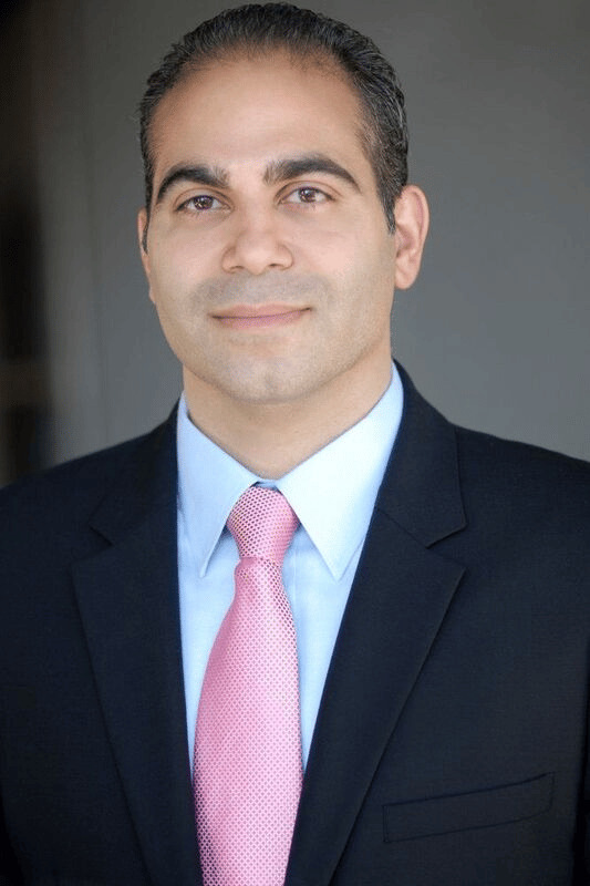
FAQ
What is an intestinal stoma?
An intestinal stoma is a surgically created opening that brings a segment of bowel through the abdominal wall to divert stool. Functionally it acts as a new organ on the abdominal surface that the patient manages daily.Why is stoma site selection done before surgery?
A site that looks ideal on an anesthetized, supine patient can vanish into a skin fold or fall on the beltline once the patient stands, sits, or bends. Marking beforehand — ideally with a WOCN — accounts for those dynamic changes and preserves a clear, visible, pouchable site with a two-inch perimeter of intact skin.How is stoma ischemia assessed?
Intraoperatively, ICG fluorescence gives a real-time map of the microvascular bed and detects early venous congestion the eye misses. At the bedside, a lubricated clear glass test tube with a penlight acts as a 360-degree speculum to trace mucosal viability and distinguish superficial from sub-fascial ischemia.Why does WHO oral rehydration solution contain sugar if patients are told to avoid sweets?
Water absorption in the small bowel depends on the SGLT-1 transporter, which moves sodium and glucose together in a fixed ratio; water then follows. The glucose in the solution isn't for calories — it's the biochemical partner that activates the sodium pump. A plain sugary drink, lacking sodium, instead pulls water into the lumen and worsens output.When can a stoma be reversed?
The traditional teaching was to wait 8–12 weeks. The Scandinavian EASY trial showed early closure of loop ileostomies at roughly 8–13 days can be as safe in carefully selected patients — though nearly 70% of patients were excluded from that trial, so selection is critical.Why are stapled reversals often preferred over hand-sewn?
The HASTA trial found comparable leak rates, so guidelines accept both. Stapled side-to-side anastomoses create a much wider common channel that stays patent even when postoperative tissue swelling peaks, so they obstruct less often in the early period.Medical Disclaimer
Colorectal Surgery Review is an educational program for clinicians and trainees and is not medical advice. Clinical decisions should be individualized and made in accordance with current guidelines and the judgment of the treating team.Keep exploring